• 文献检索
  • 文档翻译
  • 深度研究
  • 学术资讯
  • Suppr Zotero 插件Zotero 插件
  • 邀请有礼
  • 套餐&价格
  • 历史记录
应用&插件
Suppr Zotero 插件Zotero 插件浏览器插件Mac 客户端Windows 客户端微信小程序
定价
高级版会员购买积分包购买API积分包
服务
文献检索文档翻译深度研究API 文档MCP 服务
关于我们
关于 Suppr公司介绍联系我们用户协议隐私条款
关注我们

Suppr 超能文献

核心技术专利:CN118964589B侵权必究
粤ICP备2023148730 号-1Suppr @ 2026

文献检索

告别复杂PubMed语法,用中文像聊天一样搜索,搜遍4000万医学文献。AI智能推荐,让科研检索更轻松。

立即免费搜索

文件翻译

保留排版,准确专业,支持PDF/Word/PPT等文件格式,支持 12+语言互译。

免费翻译文档

深度研究

AI帮你快速写综述,25分钟生成高质量综述,智能提取关键信息,辅助科研写作。

立即免费体验

鱼类表皮角质形成细胞中肌动蛋白-肌球蛋白II系统的分析:细胞体转运机制

Analysis of the actin-myosin II system in fish epidermal keratocytes: mechanism of cell body translocation.

作者信息

Svitkina T M, Verkhovsky A B, McQuade K M, Borisy G G

机构信息

Laboratory of Molecular Biology, University of Wisconsin, Madison, Wisconsin 53706, USA.

出版信息

J Cell Biol. 1997 Oct 20;139(2):397-415. doi: 10.1083/jcb.139.2.397.

DOI:10.1083/jcb.139.2.397
PMID:9334344
原文链接:https://pmc.ncbi.nlm.nih.gov/articles/PMC2139803/
Abstract

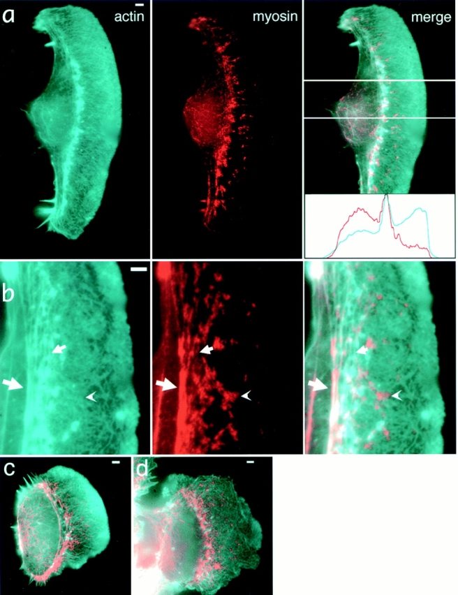
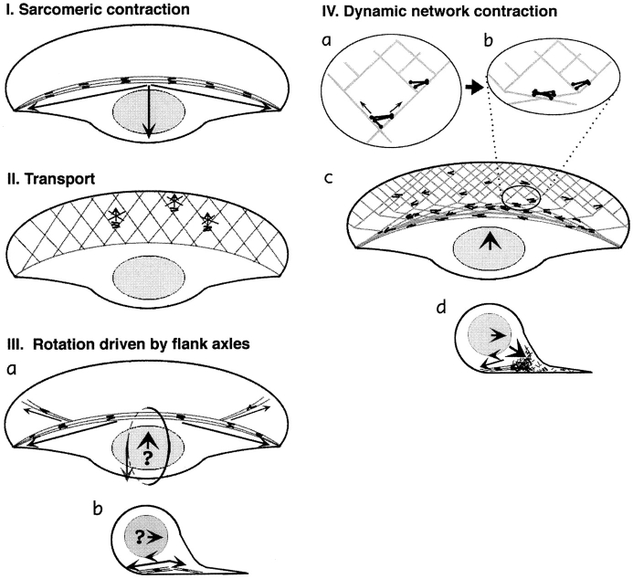
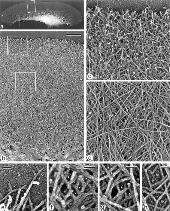
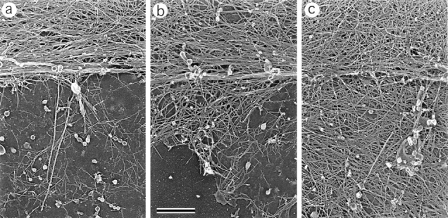
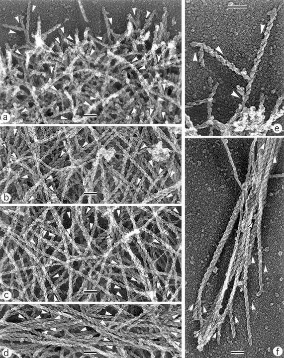
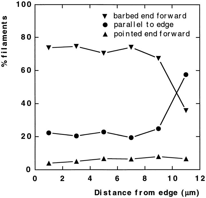
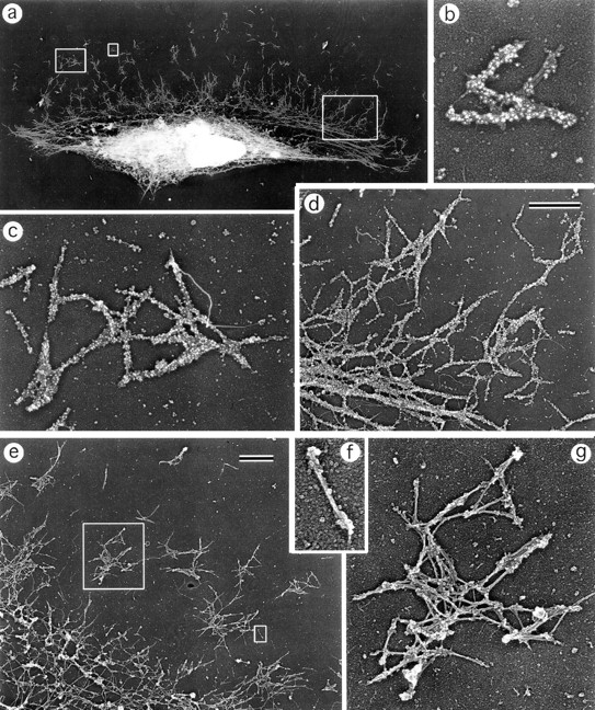
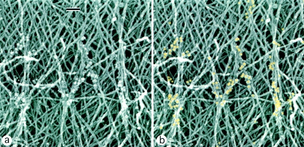
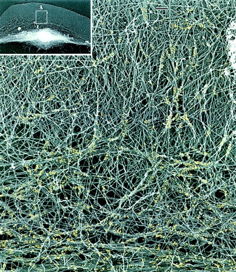
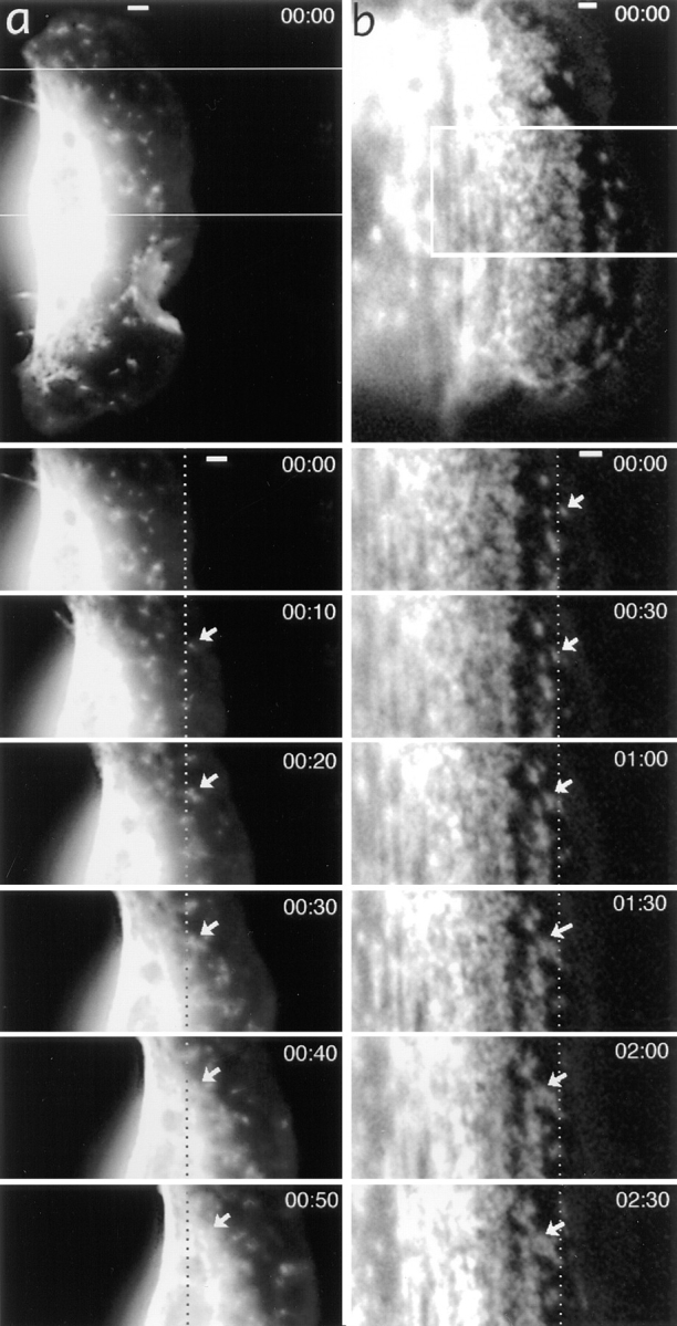
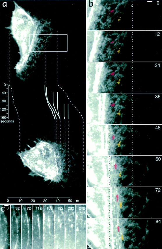
While the protrusive event of cell locomotion is thought to be driven by actin polymerization, the mechanism of forward translocation of the cell body is unclear. To elucidate the mechanism of cell body translocation, we analyzed the supramolecular organization of the actin-myosin II system and the dynamics of myosin II in fish epidermal keratocytes. In lamellipodia, long actin filaments formed dense networks with numerous free ends in a brushlike manner near the leading edge. Shorter actin filaments often formed T junctions with longer filaments in the brushlike area, suggesting that new filaments could be nucleated at sides of preexisting filaments or linked to them immediately after nucleation. The polarity of actin filaments was almost uniform, with barbed ends forward throughout most of the lamellipodia but mixed in arc-shaped filament bundles at the lamellipodial/cell body boundary. Myosin II formed discrete clusters of bipolar minifilaments in lamellipodia that increased in size and density towards the cell body boundary and colocalized with actin in boundary bundles. Time-lapse observation demonstrated that myosin clusters appeared in the lamellipodia and remained stationary with respect to the substratum in locomoting cells, but they exhibited retrograde flow in cells tethered in epithelioid colonies. Consequently, both in locomoting and stationary cells, myosin clusters approached the cell body boundary, where they became compressed and aligned, resulting in the formation of boundary bundles. In locomoting cells, the compression was associated with forward displacement of myosin features. These data are not consistent with either sarcomeric or polarized transport mechanisms of cell body translocation. We propose that the forward translocation of the cell body and retrograde flow in the lamellipodia are both driven by contraction of an actin-myosin network in the lamellipodial/cell body transition zone.

摘要

虽然细胞运动的突出事件被认为是由肌动蛋白聚合驱动的,但细胞体向前移位的机制尚不清楚。为了阐明细胞体移位的机制,我们分析了鱼类表皮角质形成细胞中肌动蛋白-肌球蛋白II系统的超分子组织和肌球蛋白II的动力学。在片状伪足中,长肌动蛋白丝在靠近前沿的位置以刷状方式形成密集网络,有许多自由末端。较短的肌动蛋白丝在刷状区域常与较长的肌动蛋白丝形成T形连接,这表明新的肌动蛋白丝可能在现有肌动蛋白丝的侧面成核,或者在成核后立即与它们相连。肌动蛋白丝的极性几乎是均匀的,在大多数片状伪足中,带刺末端向前,但在片状伪足/细胞体边界处的弧形丝束中则是混合的。肌球蛋白II在片状伪足中形成双极微丝的离散簇,其大小和密度朝着细胞体边界增加,并与边界束中的肌动蛋白共定位。延时观察表明,肌球蛋白簇出现在片状伪足中,在运动细胞中相对于基质保持静止,但在附着于上皮样菌落的细胞中它们表现出逆行流动。因此,在运动细胞和静止细胞中,肌球蛋白簇都接近细胞体边界,在那里它们被压缩并排列,导致边界束的形成。在运动细胞中,压缩与肌球蛋白特征的向前位移有关。这些数据与细胞体移位的肌节或极化运输机制均不一致。我们提出,细胞体的向前移位和片状伪足中的逆行流动都是由片状伪足/细胞体过渡区的肌动蛋白-肌球蛋白网络收缩驱动的。

https://cdn.ncbi.nlm.nih.gov/pmc/blobs/2aee/2139803/667ad6a56de7/JCB.32870f11.jpg
https://cdn.ncbi.nlm.nih.gov/pmc/blobs/2aee/2139803/58cd84849752/JCB.32870f1.jpg
https://cdn.ncbi.nlm.nih.gov/pmc/blobs/2aee/2139803/2f43ded38b19/JCB.32870f2a.jpg
https://cdn.ncbi.nlm.nih.gov/pmc/blobs/2aee/2139803/ffdf7d02deb0/JCB.32870f3a.jpg
https://cdn.ncbi.nlm.nih.gov/pmc/blobs/2aee/2139803/06b56d5cf865/JCB.32870f4a.jpg
https://cdn.ncbi.nlm.nih.gov/pmc/blobs/2aee/2139803/7072e949a125/JCB.32870f5.jpg
https://cdn.ncbi.nlm.nih.gov/pmc/blobs/2aee/2139803/4a7d73453282/JCB.32870f6a.jpg
https://cdn.ncbi.nlm.nih.gov/pmc/blobs/2aee/2139803/7500a0166138/JCB.32870f7.jpg
https://cdn.ncbi.nlm.nih.gov/pmc/blobs/2aee/2139803/0fe11fe63dde/JCB.32870f8.jpg
https://cdn.ncbi.nlm.nih.gov/pmc/blobs/2aee/2139803/4b2bda399abf/JCB.32870f9a.jpg
https://cdn.ncbi.nlm.nih.gov/pmc/blobs/2aee/2139803/77a8e61b435f/JCB.32870f10a.jpg
https://cdn.ncbi.nlm.nih.gov/pmc/blobs/2aee/2139803/667ad6a56de7/JCB.32870f11.jpg

相似文献

1
Analysis of the actin-myosin II system in fish epidermal keratocytes: mechanism of cell body translocation.鱼类表皮角质形成细胞中肌动蛋白-肌球蛋白II系统的分析:细胞体转运机制
J Cell Biol. 1997 Oct 20;139(2):397-415. doi: 10.1083/jcb.139.2.397.
2
Organization and polarity of actin filament networks in cells: implications for the mechanism of myosin-based cell motility.细胞中肌动蛋白丝网络的组织与极性:对基于肌球蛋白的细胞运动机制的启示
Biochem Soc Symp. 1999;65:173-205.
3
Identification of novel graded polarity actin filament bundles in locomoting heart fibroblasts: implications for the generation of motile force.在运动的心脏成纤维细胞中鉴定新型分级极性肌动蛋白丝束:对产生运动力的影响。
J Cell Biol. 1997 Mar 24;136(6):1287-305. doi: 10.1083/jcb.136.6.1287.
4
Nerve growth cone lamellipodia contain two populations of actin filaments that differ in organization and polarity.神经生长锥片状伪足包含两类肌动蛋白丝,它们在组织和极性上存在差异。
J Cell Biol. 1992 Dec;119(5):1219-43. doi: 10.1083/jcb.119.5.1219.
5
Polarity sorting of actin filaments in cytochalasin-treated fibroblasts.细胞松弛素处理的成纤维细胞中肌动蛋白丝的极性分选
J Cell Sci. 1997 Aug;110 ( Pt 15):1693-704. doi: 10.1242/jcs.110.15.1693.
6
Actin filament organization in the fish keratocyte lamellipodium.鱼类角膜细胞片状伪足中的肌动蛋白丝组织
J Cell Biol. 1995 Jun;129(5):1275-86. doi: 10.1083/jcb.129.5.1275.
7
Self-polarization and directional motility of cytoplasm.细胞质的自极化与定向运动性
Curr Biol. 1999 Jan 14;9(1):11-20. doi: 10.1016/s0960-9822(99)80042-6.
8
Myosin II filament assemblies in the active lamella of fibroblasts: their morphogenesis and role in the formation of actin filament bundles.成纤维细胞活跃薄片中的肌球蛋白II丝组装体:它们的形态发生及其在肌动蛋白丝束形成中的作用。
J Cell Biol. 1995 Nov;131(4):989-1002. doi: 10.1083/jcb.131.4.989.
9
Network contraction model for cell translocation and retrograde flow.用于细胞迁移和顺行流动的网络收缩模型。
Biochem Soc Symp. 1999;65:207-22.
10
Polarity sorting drives remodeling of actin-myosin networks.极性排序驱动肌动球蛋白网络的重塑。
J Cell Sci. 2018 Dec 13;132(4):jcs219717. doi: 10.1242/jcs.219717.

引用本文的文献

1
Myosin 2 drives actin contractility in fast-crawling species outside of the amorphean lineage.肌球蛋白2在无变形虫谱系之外的快速爬行物种中驱动肌动蛋白收缩。
bioRxiv. 2025 May 19:2025.05.16.654244. doi: 10.1101/2025.05.16.654244.
2
Cryo-ET of actin cytoskeleton and membrane structure in lamellipodia formation using optogenetics.利用光遗传学对片足形成过程中的肌动蛋白细胞骨架和膜结构进行冷冻电子断层扫描。
iScience. 2025 Apr 24;28(6):112529. doi: 10.1016/j.isci.2025.112529. eCollection 2025 Jun 20.
3
Endogenous SH2B1 protein localizes to lamellipodia and filopodia: platinum replica electron-microscopy study.

本文引用的文献

1
The nucleation-release model of actin filament dynamics in cell motility.细胞运动中肌动蛋白丝动力学的成核-释放模型。
Trends Cell Biol. 1992 Aug;2(8):219-22. doi: 10.1016/0962-8924(92)90298-2.
2
ELECTRON MICROSCOPE STUDIES ON THE STRUCTURE OF NATURAL AND SYNTHETIC PROTEIN FILAMENTS FROM STRIATED MUSCLE.横纹肌天然及合成蛋白细丝结构的电子显微镜研究
J Mol Biol. 1963 Sep;7:281-308. doi: 10.1016/s0022-2836(63)80008-x.
3
Polarity sorting of actin filaments in cytochalasin-treated fibroblasts.细胞松弛素处理的成纤维细胞中肌动蛋白丝的极性分选
内源性SH2B1蛋白定位于片状伪足和丝状伪足:铂复型电子显微镜研究。
MicroPubl Biol. 2025 Jan 17;2025. doi: 10.17912/micropub.biology.001451. eCollection 2025.
4
A simple active fluid model unites cytokinesis, cell crawling, and axonal outgrowth.一个简单的活性流体模型将胞质分裂、细胞爬行和轴突生长统一起来。
Front Cell Dev Biol. 2024 Oct 17;12:1491429. doi: 10.3389/fcell.2024.1491429. eCollection 2024.
5
Non-Muscle Myosin II A: Friend or Foe in Cancer?非肌肉肌球蛋白 IIA:癌症的朋友还是敌人?
Int J Mol Sci. 2024 Aug 30;25(17):9435. doi: 10.3390/ijms25179435.
6
Cell blebbing novel therapeutic possibilities to counter metastasis.细胞起泡为对抗转移提供了新的治疗可能性。
Clin Exp Metastasis. 2024 Dec;41(6):817-828. doi: 10.1007/s10585-024-10308-z. Epub 2024 Sep 2.
7
Myosin forces elicit an F-actin structural landscape that mediates mechanosensitive protein recognition.肌球蛋白力引发一种F-肌动蛋白结构格局,该格局介导机械敏感蛋白识别。
bioRxiv. 2024 Aug 17:2024.08.15.608188. doi: 10.1101/2024.08.15.608188.
8
Structure, regulation, and mechanisms of nonmuscle myosin-2.非肌肉肌球蛋白-2 的结构、调控和机制。
Cell Mol Life Sci. 2024 Jun 15;81(1):263. doi: 10.1007/s00018-024-05264-6.
9
A simple active fluid model unites cytokinesis, cell crawling, and axonal outgrowth.一个简单的活性流体模型将胞质分裂、细胞爬行和轴突生长统一起来。
bioRxiv. 2024 May 23:2024.05.22.595337. doi: 10.1101/2024.05.22.595337.
10
Coordination of actin plus-end dynamics by IQGAP1, formin, and capping protein.IQGAP1、成核因子和加帽蛋白协调肌动蛋白末端动力学。
J Cell Biol. 2024 Sep 2;223(9). doi: 10.1083/jcb.202305065. Epub 2024 May 24.
J Cell Sci. 1997 Aug;110 ( Pt 15):1693-704. doi: 10.1242/jcs.110.15.1693.
4
Cell motility: complex dynamics at the leading edge.细胞运动性:前沿的复杂动力学
Curr Biol. 1997 Mar 1;7(3):R164-7. doi: 10.1016/s0960-9822(97)70079-4.
5
Xenopus actin depolymerizing factor/cofilin (XAC) is responsible for the turnover of actin filaments in Listeria monocytogenes tails.非洲爪蟾肌动蛋白解聚因子/丝切蛋白(XAC)负责单核细胞增生李斯特菌尾部肌动蛋白丝的周转。
J Cell Biol. 1997 Mar 24;136(6):1323-32. doi: 10.1083/jcb.136.6.1323.
6
Actin depolymerizing factor (ADF/cofilin) enhances the rate of filament turnover: implication in actin-based motility.肌动蛋白解聚因子(ADF/丝切蛋白)提高了丝状体周转的速率:对基于肌动蛋白的运动的影响。
J Cell Biol. 1997 Mar 24;136(6):1307-22. doi: 10.1083/jcb.136.6.1307.
7
Identification of novel graded polarity actin filament bundles in locomoting heart fibroblasts: implications for the generation of motile force.在运动的心脏成纤维细胞中鉴定新型分级极性肌动蛋白丝束:对产生运动力的影响。
J Cell Biol. 1997 Mar 24;136(6):1287-305. doi: 10.1083/jcb.136.6.1287.
8
Accelerating on a treadmill: ADF/cofilin promotes rapid actin filament turnover in the dynamic cytoskeleton.在跑步机上加速:ADF/丝切蛋白促进动态细胞骨架中肌动蛋白丝的快速周转。
J Cell Biol. 1997 Mar 24;136(6):1165-8. doi: 10.1083/jcb.136.6.1165.
9
Structure, subunit topology, and actin-binding activity of the Arp2/3 complex from Acanthamoeba.棘阿米巴属中Arp2/3复合物的结构、亚基拓扑结构及肌动蛋白结合活性
J Cell Biol. 1997 Jan 27;136(2):331-43. doi: 10.1083/jcb.136.2.331.
10
Actin polymerization is induced by Arp2/3 protein complex at the surface of Listeria monocytogenes.肌动蛋白聚合由单核细胞增生李斯特菌表面的Arp2/3蛋白复合物诱导。
Nature. 1997 Jan 16;385(6613):265-9. doi: 10.1038/385265a0.