Matsuda H, Koyama H, Sato H, Sawada J, Itakura A, Tanaka A, Matsumoto M, Konno K, Ushio H, Matsuda K
Department of Veterinary Clinic, Faculty of Agriculture, Tokyo University of Agriculture and Technology, Fuchu, Tokyo 183, Japan.
J Exp Med. 1998 Feb 2;187(3):297-306. doi: 10.1084/jem.187.3.297.
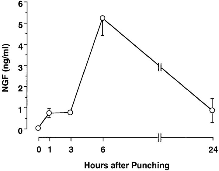
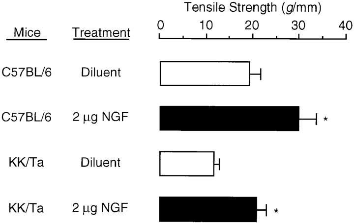
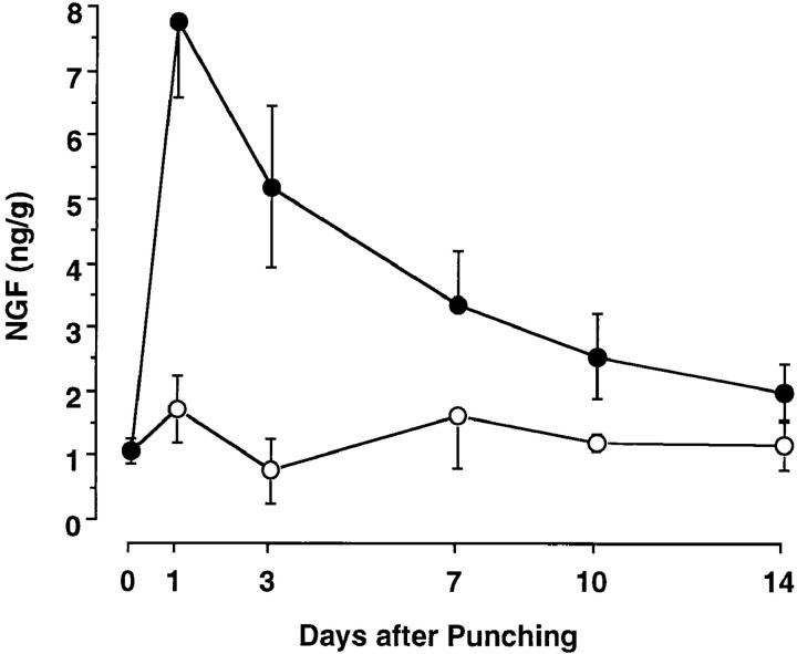
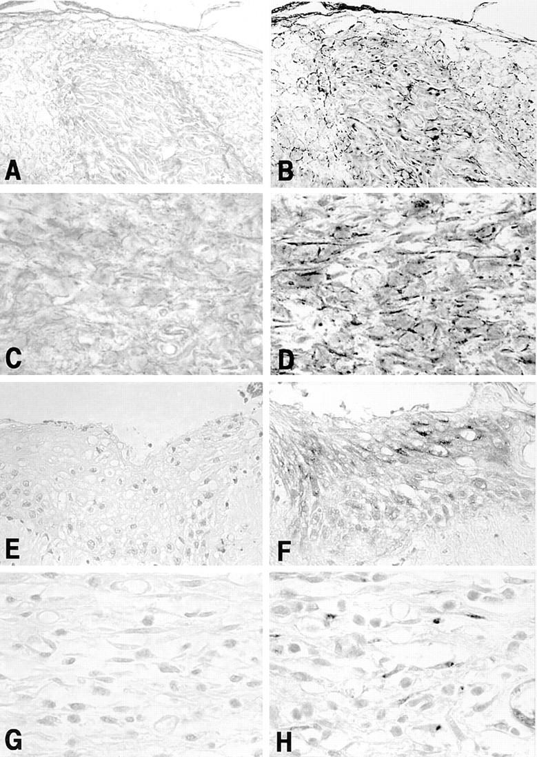
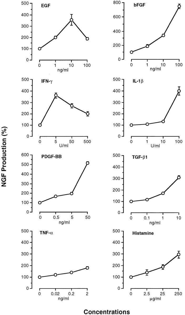
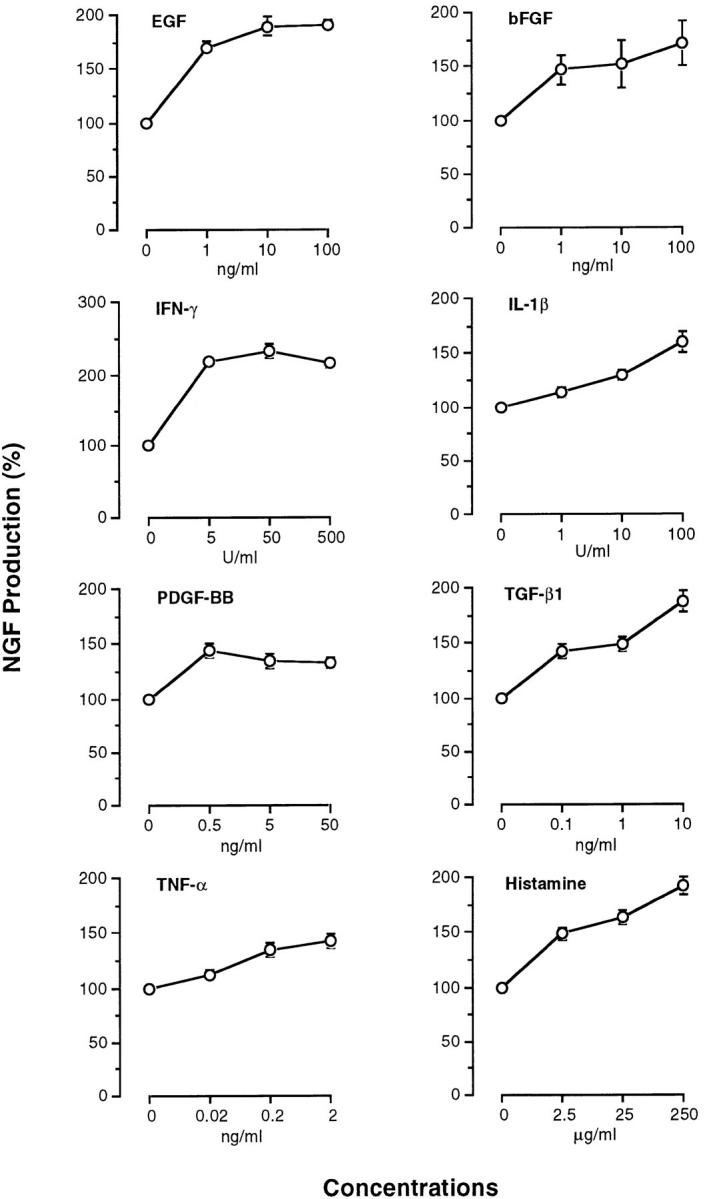
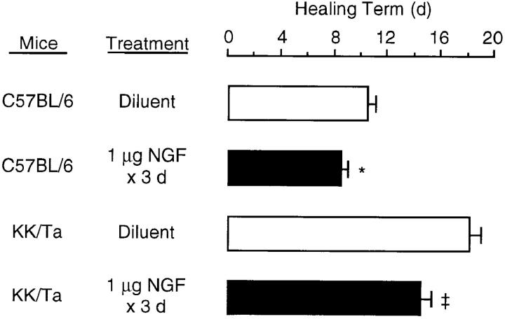
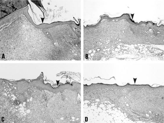
Four full-thickness skin wounds made in normal mice led to the significant increase in levels of nerve growth factor (NGF) in sera and in wounded skin tissues. Since sialoadenectomy before the wounds inhibited the rise in serum levels of NGF, the NGF may be released from the salivary gland into the blood stream after the wounds. In contrast, the fact that messenger RNA and protein of NGF were detected in newly formed epithelial cells at the edge of the wound and fibroblasts consistent with the granulation tissue produced in the wound space, suggests that NGF was also produced at the wounded skin site. Topical application of NGF into the wounds accelerated the rate of wound healing in normal mice and in healing-impaired diabetic KK/Ta mice. This clinical effect of NGF was evaluated by histological examination; the increases in the degree of reepithelialization, the thickness of the granulation tissue, and the density of extracellular matrix were observed. NGF also increased the breaking strength of healing linear wounds in normal and diabetic mice. These findings suggested that NGF immediately and constitutively released in response to cutaneous injury may contribute to wound healing through broader biological activities, and NGF improved the diabetic impaired response of wound healing.
在正常小鼠身上制造的四处全层皮肤伤口导致血清和受伤皮肤组织中神经生长因子(NGF)水平显著升高。由于伤口形成前进行涎腺切除术可抑制血清中NGF水平的升高,因此NGF可能在伤口形成后从唾液腺释放到血流中。相反,在伤口边缘新形成的上皮细胞和成纤维细胞中检测到与伤口空间中产生的肉芽组织一致的NGF信使核糖核酸和蛋白质,这表明受伤皮肤部位也产生NGF。将NGF局部应用于伤口可加速正常小鼠和愈合受损的糖尿病KK/Ta小鼠的伤口愈合速度。NGF的这种临床效果通过组织学检查进行评估;观察到再上皮化程度、肉芽组织厚度和细胞外基质密度增加。NGF还提高了正常和糖尿病小鼠愈合线性伤口的抗张强度。这些发现表明,对皮肤损伤立即产生并持续释放的NGF可能通过更广泛的生物学活性促进伤口愈合,并且NGF改善了糖尿病患者受损的伤口愈合反应。