Doglioni C, De Boni M, Cielo R, Laurino L, Pelosio P, Braidotti P, Viale G
Department of Surgical Pathology, Ospedale Civile, Feltre, Italy.
J Clin Pathol. 1992 Nov;45(11):964-7. doi: 10.1136/jcp.45.11.964.
To assess the prevalence of gastric giardiasis in patients undergoing upper gastrointestinal endoscopy, and to define the clinicopathological correlates of gastric Giardia lamblia infection.
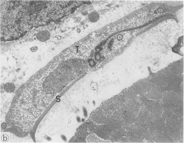
Consecutive gastric biopsy specimens (n = 15,023) from 11,085 patients, taken at Feltre City Hospital (north eastern Italy) from January 1986 to December 1991, were histologically and immunocytochemically examined for the occurrence of G lamblia trophozoites. Three gastric biopsy specimens from patients harbouring G lamblia infection, who repeated endoscopy before treatment, were also examined electron microscopically.
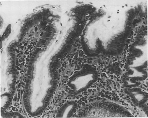
Forty one patients (0.37% of the population study) harboured gastric giardiasis. All patients underwent upper gastrointestinal endoscopy because of dyspepsia, epigastric pain, or abdominal distension. Only two patients had diarrhoea at the time of investigation. Giardiasis was clinically unsuspected in all cases, although the nine patients who also had duodenal biopsies performed had concomitant intestinal giardiasis. Gastric giardiasis was invariably associated with chronic atrophic gastritis. Intestinal metaplasia of the gastric mucosa and Helicobacter pylori infection were found in 32 and 37 of the 41 patients with gastric giardiasis, respectively.
The invariable association of gastric giardiasis with chronic atrophic gastritis, most often showing intestinal metaplasia and H pylori infection, indicates that a decreased gastric acidity is a prerequisite for localisation of G lamblia to the gastric mucosa. Though its possible role as a gastric pathogen remains to be elucidated, these findings suggest that trophozoites should be carefully searched for when examining gastric biopsy specimens showing chronic atrophic gastritis.
评估接受上消化道内镜检查患者中胃贾第虫病的患病率,并确定胃蓝氏贾第鞭毛虫感染的临床病理相关性。
对1986年1月至1991年12月在意大利东北部费尔特雷市医院采集的11,085例患者的连续胃活检标本(n = 15,023)进行组织学和免疫细胞化学检查,以检测蓝氏贾第鞭毛虫滋养体的存在。对3例在治疗前重复进行内镜检查的蓝氏贾第鞭毛虫感染患者的胃活检标本也进行了电子显微镜检查。
41例患者(占研究人群的0.37%)患有胃贾第虫病。所有患者均因消化不良、上腹部疼痛或腹胀接受上消化道内镜检查。在调查时只有2例患者有腹泻。所有病例临床上均未怀疑有贾第虫病,尽管9例同时进行十二指肠活检的患者伴有肠道贾第虫病。胃贾第虫病总是与慢性萎缩性胃炎相关。41例胃贾第虫病患者中,分别有32例和37例发现胃黏膜肠化生和幽门螺杆菌感染。
胃贾第虫病总是与慢性萎缩性胃炎相关,后者最常表现为肠化生和幽门螺杆菌感染,这表明胃酸降低是蓝氏贾第鞭毛虫定位于胃黏膜的先决条件。尽管其作为胃病原体的可能作用仍有待阐明,但这些发现表明,在检查显示慢性萎缩性胃炎的胃活检标本时,应仔细寻找滋养体。