• 文献检索
  • 文档翻译
  • 深度研究
  • 学术资讯
  • Suppr Zotero 插件Zotero 插件
  • 邀请有礼
  • 套餐&价格
  • 历史记录
应用&插件
Suppr Zotero 插件Zotero 插件浏览器插件Mac 客户端Windows 客户端微信小程序
定价
高级版会员购买积分包购买API积分包
服务
文献检索文档翻译深度研究API 文档MCP 服务
关于我们
关于 Suppr公司介绍联系我们用户协议隐私条款
关注我们

Suppr 超能文献

核心技术专利:CN118964589B侵权必究
粤ICP备2023148730 号-1Suppr @ 2026

文献检索

告别复杂PubMed语法,用中文像聊天一样搜索,搜遍4000万医学文献。AI智能推荐,让科研检索更轻松。

立即免费搜索

文件翻译

保留排版,准确专业,支持PDF/Word/PPT等文件格式,支持 12+语言互译。

免费翻译文档

深度研究

AI帮你快速写综述,25分钟生成高质量综述,智能提取关键信息,辅助科研写作。

立即免费体验

用于重建三维组织纳米结构的连续块面扫描电子显微镜技术。

Serial block-face scanning electron microscopy to reconstruct three-dimensional tissue nanostructure.

作者信息

Denk Winfried, Horstmann Heinz

机构信息

Max Planck Institute for Medical Research, Heidelberg, Germany.

出版信息

PLoS Biol. 2004 Nov;2(11):e329. doi: 10.1371/journal.pbio.0020329. Epub 2004 Oct 19.

DOI:10.1371/journal.pbio.0020329
PMID:15514700
原文链接:https://pmc.ncbi.nlm.nih.gov/articles/PMC524270/
Abstract

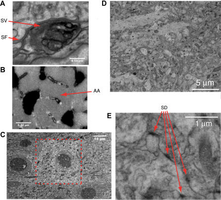
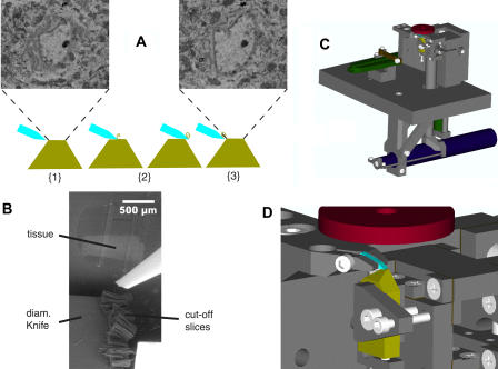
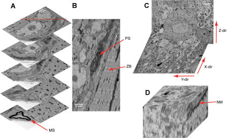
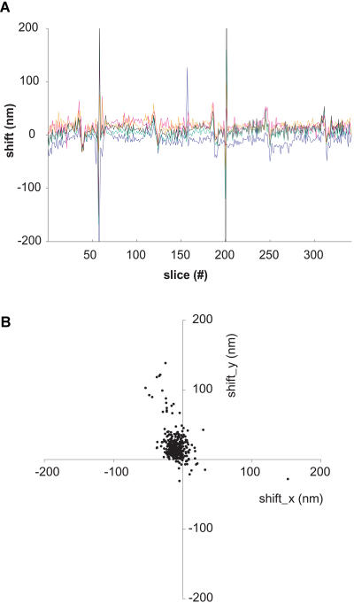
Three-dimensional (3D) structural information on many length scales is of central importance in biological research. Excellent methods exist to obtain structures of molecules at atomic, organelles at electron microscopic, and tissue at light-microscopic resolution. A gap exists, however, when 3D tissue structure needs to be reconstructed over hundreds of micrometers with a resolution sufficient to follow the thinnest cellular processes and to identify small organelles such as synaptic vesicles. Such 3D data are, however, essential to understand cellular networks that, particularly in the nervous system, need to be completely reconstructed throughout a substantial spatial volume. Here we demonstrate that datasets meeting these requirements can be obtained by automated block-face imaging combined with serial sectioning inside the chamber of a scanning electron microscope. Backscattering contrast is used to visualize the heavy-metal staining of tissue prepared using techniques that are routine for transmission electron microscopy. Low-vacuum (20-60 Pa H(2)O) conditions prevent charging of the uncoated block face. The resolution is sufficient to trace even the thinnest axons and to identify synapses. Stacks of several hundred sections, 50-70 nm thick, have been obtained at a lateral position jitter of typically under 10 nm. This opens the possibility of automatically obtaining the electron-microscope-level 3D datasets needed to completely reconstruct the connectivity of neuronal circuits.

摘要

在许多长度尺度上的三维(3D)结构信息在生物学研究中至关重要。现已有出色的方法来获取原子水平的分子结构、电子显微镜水平的细胞器结构以及光学显微镜水平的组织结构。然而,当需要在数百微米范围内重建三维组织结构,且分辨率要足以追踪最细的细胞突起并识别诸如突触小泡等小细胞器时,就存在差距。不过,此类三维数据对于理解细胞网络至关重要,尤其是在神经系统中,需要在相当大的空间体积内完整重建细胞网络。在此,我们证明通过在扫描电子显微镜腔室内结合连续切片进行自动块面成像,可以获得满足这些要求的数据集。背散射对比度用于可视化使用透射电子显微镜常规技术制备的组织的重金属染色。低真空(20 - 60 Pa H₂O)条件可防止未镀膜块面带电。分辨率足以追踪最细的轴突并识别突触。已获得数百个厚度为50 - 70 nm的切片堆栈,其横向位置抖动通常在10 nm以下。这开启了自动获取完全重建神经元回路连接所需的电子显微镜水平三维数据集的可能性。

https://cdn.ncbi.nlm.nih.gov/pmc/blobs/65c5/524270/77b1ebfce15b/pbio.0020329.g005.jpg
https://cdn.ncbi.nlm.nih.gov/pmc/blobs/65c5/524270/e8555fa5f4f3/pbio.0020329.g001.jpg
https://cdn.ncbi.nlm.nih.gov/pmc/blobs/65c5/524270/a771a3e523bc/pbio.0020329.g002.jpg
https://cdn.ncbi.nlm.nih.gov/pmc/blobs/65c5/524270/061a343f4340/pbio.0020329.g003.jpg
https://cdn.ncbi.nlm.nih.gov/pmc/blobs/65c5/524270/0b5a3bc64093/pbio.0020329.g004.jpg
https://cdn.ncbi.nlm.nih.gov/pmc/blobs/65c5/524270/77b1ebfce15b/pbio.0020329.g005.jpg

相似文献

1
Serial block-face scanning electron microscopy to reconstruct three-dimensional tissue nanostructure.用于重建三维组织纳米结构的连续块面扫描电子显微镜技术。
PLoS Biol. 2004 Nov;2(11):e329. doi: 10.1371/journal.pbio.0020329. Epub 2004 Oct 19.
2
Developing 3D SEM in a broad biological context.在广泛的生物学背景下开发三维扫描电子显微镜。
J Microsc. 2015 Aug;259(2):80-96. doi: 10.1111/jmi.12211. Epub 2015 Jan 26.
3
Knife-edge scanning microscopy for imaging and reconstruction of three-dimensional anatomical structures of the mouse brain.用于小鼠大脑三维解剖结构成像与重建的刀口扫描显微镜。
J Microsc. 2008 Jul;231(Pt 1):134-43. doi: 10.1111/j.1365-2818.2008.02024.x.
4
Combining serial block face and focused ion beam scanning electron microscopy for 3D studies of rare events.结合连续块面和聚焦离子束扫描电子显微镜用于罕见事件的三维研究。
Methods Cell Biol. 2019;152:87-101. doi: 10.1016/bs.mcb.2019.03.014. Epub 2019 May 7.
5
New developments in electron microscopy for serial image acquisition of neuronal profiles.用于神经元轮廓连续图像采集的电子显微镜新进展。
Microscopy (Oxf). 2015 Feb;64(1):27-36. doi: 10.1093/jmicro/dfu111. Epub 2015 Jan 5.
6
Towards the imaging of Weibel-Palade body biogenesis by serial block face-scanning electron microscopy.通过连续块面扫描电子显微镜对魏贝尔-帕拉德小体生物发生进行成像研究。
J Microsc. 2015 Aug;259(2):97-104. doi: 10.1111/jmi.12222. Epub 2015 Jan 23.
7
Contour-propagation algorithms for semi-automated reconstruction of neural processes.用于神经突起半自动重建的轮廓传播算法。
J Neurosci Methods. 2008 Jan 30;167(2):349-57. doi: 10.1016/j.jneumeth.2007.07.021. Epub 2007 Aug 10.
8
Rapid specimen preparation to improve the throughput of electron microscopic volume imaging for three-dimensional analyses of subcellular ultrastructures with serial block-face scanning electron microscopy.快速标本制备以提高用于亚细胞超微结构三维分析的电子显微镜体积成像的通量,采用连续块面扫描电子显微镜技术。
Med Mol Morphol. 2016 Sep;49(3):154-62. doi: 10.1007/s00795-016-0134-7. Epub 2016 Feb 11.
9
Challenges of microtome-based serial block-face scanning electron microscopy in neuroscience.基于切片机的连续块面扫描电子显微镜在神经科学中的挑战。
J Microsc. 2015 Aug;259(2):137-142. doi: 10.1111/jmi.12244. Epub 2015 Apr 23.
10
Optimizing the 3D-reconstruction technique for serial block-face scanning electron microscopy.优化用于连续块面扫描电子显微镜的三维重建技术。
J Neurosci Methods. 2016 May 1;264:16-24. doi: 10.1016/j.jneumeth.2016.02.019. Epub 2016 Feb 27.

引用本文的文献

1
Cutting-edge technologies in neural regeneration.神经再生领域的前沿技术。
Cell Regen. 2025 Sep 5;14(1):38. doi: 10.1186/s13619-025-00260-y.
2
An Optimized Protocol for SBEM-Based Ultrastructural Analysis of Cultured Human Cells.一种基于扫描电子显微镜断层成像技术的培养人细胞超微结构分析优化方案
Methods Protoc. 2025 Aug 6;8(4):90. doi: 10.3390/mps8040090.
3
Three-dimensional ultrastructural differences between thalamic and non-thalamic recipient layers in macaque V1.猕猴初级视皮层中丘脑和非丘脑接受层之间的三维超微结构差异

本文引用的文献

1
The structure of the nervous system of the nematode Caenorhabditis elegans.秀丽隐杆线虫的神经系统结构。
Philos Trans R Soc Lond B Biol Sci. 1986 Nov 12;314(1165):1-340. doi: 10.1098/rstb.1986.0056.
2
RETINAL GANGLION CELLS RESPONDING SELECTIVELY TO DIRECTION AND SPEED OF IMAGE MOTION IN THE RABBIT.兔视网膜神经节细胞对图像运动的方向和速度具有选择性反应。
J Physiol. 1964 Oct;173(3):377-407. doi: 10.1113/jphysiol.1964.sp007463.
3
AN IMPROVED STAINING METHOD FOR ELECTRON MICROSCOPY.一种改进的电子显微镜染色方法。
bioRxiv. 2025 Aug 6:2025.08.04.668334. doi: 10.1101/2025.08.04.668334.
4
Deep mapping of the of cerebellar Purkinje neurons.小脑浦肯野神经元的深度图谱绘制。 你提供的原文似乎不太完整,“of the”中间应该还有具体内容。以上是根据现有内容翻译的结果。
bioRxiv. 2025 Jul 14:2025.07.08.663701. doi: 10.1101/2025.07.08.663701.
5
The physical consequences of sperm gigantism.精子巨大症的生理后果。
bioRxiv. 2025 Jul 23:2025.07.19.665658. doi: 10.1101/2025.07.19.665658.
6
PyReconstruct: A fully open-source, collaborative successor to Reconstruct.PyReconstruct:Reconstruct的一个完全开源的协作式后续版本。
Proc Natl Acad Sci U S A. 2025 Aug 5;122(31):e2505822122. doi: 10.1073/pnas.2505822122. Epub 2025 Jul 30.
7
Biological volume EM with focused Ga ion beam depends on formation of radiation-resistant Ga-rich layer at block face.使用聚焦镓离子束的生物体积电子显微镜检查取决于在块表面形成抗辐射富镓层。
Res Sq. 2025 Jul 10:rs.3.rs-5235218. doi: 10.21203/rs.3.rs-5235218/v1.
8
A scalable, all-optical method for mapping synaptic connectivity with cell-type specificity.一种用于绘制具有细胞类型特异性的突触连接性的可扩展全光学方法。
bioRxiv. 2025 Jun 25:2025.06.25.661552. doi: 10.1101/2025.06.25.661552.
9
Volume electron microscopy reveals 3D synaptic nanoarchitecture in postmortem human prefrontal cortex.体积电子显微镜揭示了死后人类前额叶皮质中的三维突触纳米结构。
iScience. 2025 May 26;28(7):112747. doi: 10.1016/j.isci.2025.112747. eCollection 2025 Jul 18.
10
Morphology and ultrastructure of external sense organs of larvae.幼虫外部感觉器官的形态学与超微结构
Elife. 2025 Jun 16;12:RP91155. doi: 10.7554/eLife.91155.
J Cell Biol. 1964 Sep;22(3):697-701. doi: 10.1083/jcb.22.3.697.
4
Ultrastructure of retinal rod synapses of the guinea pig eye as revealed by three-dimensional reconstructions from serial sections.通过连续切片的三维重建揭示的豚鼠眼视网膜视杆突触的超微结构。
J Ultrastruct Res. 1958 Nov;2(1):122-70. doi: 10.1016/s0022-5320(58)90050-9.
5
The use of environmental scanning electron microscopy for imaging wet and insulating materials.环境扫描电子显微镜在湿态和绝缘材料成像中的应用。
Nat Mater. 2003 Aug;2(8):511-6. doi: 10.1038/nmat898.
6
Electron tomography: towards visualizing the molecular organization of the cytoplasm.电子断层扫描:迈向可视化细胞质的分子组织
Curr Opin Struct Biol. 2002 Oct;12(5):679-84. doi: 10.1016/s0959-440x(02)00378-0.
7
Surface imaging microscopy, an automated method for visualizing whole embryo samples in three dimensions at high resolution.表面成像显微镜,一种用于在三维空间中以高分辨率可视化整个胚胎样本的自动化方法。
Dev Dyn. 2002 Nov;225(3):369-75. doi: 10.1002/dvdy.10169.
8
Directionally selective calcium signals in dendrites of starburst amacrine cells.星爆无长突细胞树突中的方向选择性钙信号
Nature. 2002 Aug 22;418(6900):845-52. doi: 10.1038/nature00931. Epub 2002 Aug 4.
9
Quantitative serial sectioning analysis.定量连续切片分析
J Microsc. 2001 Mar;201(Pt 3):388-94. doi: 10.1046/j.1365-2818.2001.00832.x.
10
Imaging neuronal subsets in transgenic mice expressing multiple spectral variants of GFP.对表达多种绿色荧光蛋白光谱变体的转基因小鼠中的神经元亚群进行成像。
Neuron. 2000 Oct;28(1):41-51. doi: 10.1016/s0896-6273(00)00084-2.