Trerè D, Zilbering A, Dittus D, Kim P, Ginsberg P C, Daskal I
Albert Einstein Medical Center, 5501 Old York Road, Philadelphia, PA 19141-3098, USA.
Clin Mol Pathol. 1996 Aug;49(4):M209-13. doi: 10.1136/mp.49.4.m209.
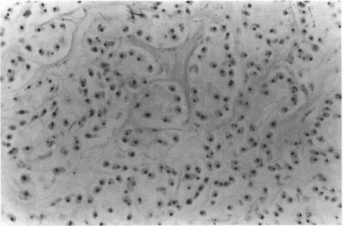
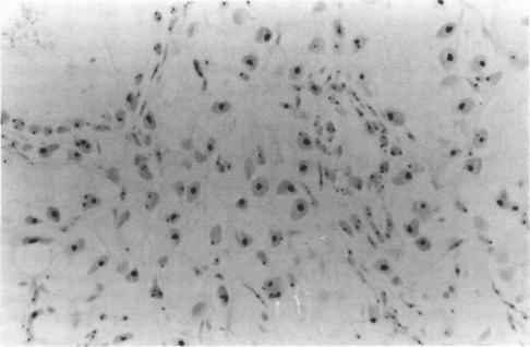
Aims-To define the relation between the quantity of silver stained nucleolar organiser regions (AgNORs) and histological grade, clinical stage, DNA content, and MIB-1 immunostaining in needle biopsy specimens of prostatic adenocarcinomas.Methods-Histological grade was determined according to the Gleason system. AgNOR quantity, DNA content and MIB-1 immunostaining were evaluated by image cytometry on routine histological sections stained with silver, Feulgen reaction and MIB-1 antibody, respectively.Results-The mean AgNOR area increased with increasing Gleason score. A significant difference was found in the AgNOR values between low, intermediate and high grade tumours. Patients with clinically localised tumour (stages A and B) had lower AgNOR values than patients with advanced disease (stages C and D), but the difference in the mean AgNOR values between the two groups was not statistically significant. Non-diploid tumours had a significantly higher mean (SD) AgNOR area than diploid tumours (3.68 (1.04) mum(2)v 2.73 (0.60) mum(2), respectively), while no significant difference was observed in the mean AgNOR values between aneuploid and tetraploid tumours (3.68 (1.04) mum(2)v 3.70 (1.05) mum(2)). When AgNOR and MIB-1-PI values were compared using linear regression analysis, a highly significant correlation was found.Conclusions-These data demonstrate that AgNOR quantity reflects the proliferative potential of prostatic adenocarcinomas, and is significantly related to histological grade and DNA content. The ease of application on routine sections, maintaining the morphological integrity of the tissue, the ability to evaluate selected histological areas of limited size and objective quantification by image cytometry make the AgNOR method particularly suitable for cell kinetic analysis in prostatic needle biopsy specimens.
目的——确定前列腺腺癌针吸活检标本中银染核仁组成区(AgNORs)数量与组织学分级、临床分期、DNA含量及MIB-1免疫染色之间的关系。方法——根据Gleason系统确定组织学分级。分别通过图像细胞术对用银染色、福尔根反应和MIB-1抗体染色的常规组织切片评估AgNOR数量、DNA含量及MIB-1免疫染色。结果——平均AgNOR面积随Gleason评分增加而增大。低级别、中级别和高级别肿瘤之间的AgNOR值存在显著差异。临床局限性肿瘤(A期和B期)患者的AgNOR值低于晚期疾病(C期和D期)患者,但两组之间平均AgNOR值的差异无统计学意义。非二倍体肿瘤的平均(标准差)AgNOR面积显著高于二倍体肿瘤(分别为3.68(1.04)μm²对2.73(0.60)μm²),而异倍体和四倍体肿瘤之间的平均AgNOR值未观察到显著差异(3.68(1.04)μm²对3.70(1.05)μm²)。使用线性回归分析比较AgNOR和MIB-1增殖指数(PI)值时,发现高度显著的相关性。结论——这些数据表明,AgNOR数量反映了前列腺腺癌的增殖潜能,且与组织学分级和DNA含量显著相关。AgNOR方法易于应用于常规切片,能保持组织的形态完整性,能够评估有限大小的选定组织学区域,并通过图像细胞术进行客观定量,这使得AgNOR方法特别适用于前列腺针吸活检标本的细胞动力学分析。