Prachasilpchai W, Nuanualsuwan S, Chatsuwan T, Techangamsuwan S, Wangnaitham S, Sailasuta A
Department of Pathology, Faculty of Veterinary Science, Chulalongkorn University, Bangkok 10330, Thailand.
J Vet Sci. 2007 Jun;8(2):139-45. doi: 10.4142/jvs.2007.8.2.139.
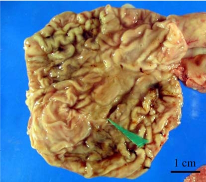
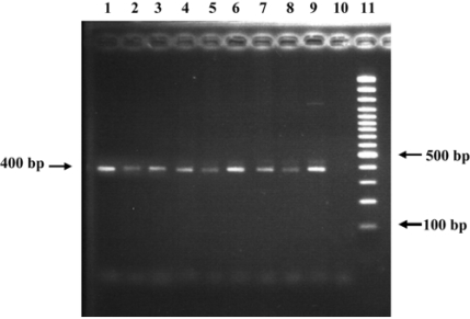
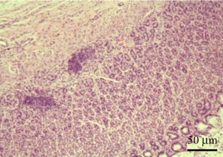
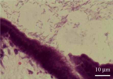
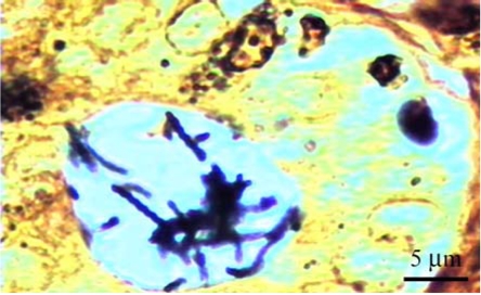
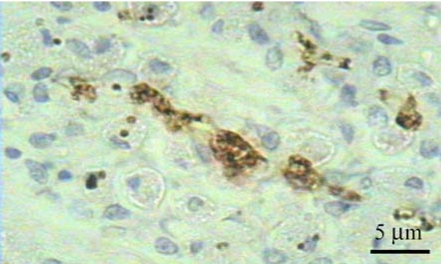
A total of 75 biopsied samples of cardia, fundus, body, and pyloric antrum from necropsied dogs that were submitted to the Department of Pathology, Faculty of Veterinary Science, Chulalongkorn University from April 2003 to June 2004 were investigated. The objectives of this study were to determine the prevalence of Helicobacter spp. in canine stomach by polymerase chain reaction (PCR) in comparison to histochemistry versus immunohistochemistry (IHC), and to correlate these diagnostic methods with the clinical significance in infected dogs. Histopathological results revealed 60.0% (45/75) of samples to be positive, and consisted of mild gastritis in 64.44% (29/45), moderate gastritis in 11.11% (5/45), and severe gastritis in 24.44% (11/45). The proportion showing no histopathological lesions was 40.0% (30/75). Helicobacter spp. were localized to the luminal crypt in 18.67% (14/75), gastric pit in 22.67% (17/75), gastric gland in 21.33% (16/75), and gastric epithelium in 8% (6/75). The percentages of positive samples of Helicobacter spp. diagnosed by hematoxylin and eosin stain (H&E), Warthin Starry stain (WSS), IHC with rabbit polyclonal anti-H. pylori antibody, and PCR were 17.3% (13/75), 46.7% (35/75), 30.7% (23/75), and 10.7% (8/75), respectively. No significant differences were observed in histopathological changes in portions of the stomach (p > 0.05). The diagnosis of Helicobacter spp. by PCR in comparison to that by WSS and IHC was not significantly different (p > 0.05). There were no relationships between pathological studies using H&E, WSS, and IHC, and especially between PCR and clinical signs of Helicobacter spp. infections in canine stomachs (p > 0.05). The present study revealed significantly different levels of correlation for Helicobacter spp. detection between H&E and WSS (p < 0.001). Results indicate that the method of choice for diagnosis of Helicobacter spp. infection in canine stomach is dependent on the purpose of study and appropriate specimen collection.
对2003年4月至2004年6月提交给朱拉隆功大学兽医学院病理学系的75份来自尸检犬的贲门、胃底、胃体和幽门窦活检样本进行了研究。本研究的目的是通过聚合酶链反应(PCR)与组织化学和免疫组织化学(IHC)比较,确定犬胃中幽门螺杆菌属的患病率,并将这些诊断方法与感染犬的临床意义相关联。组织病理学结果显示60.0%(45/75)的样本呈阳性,其中轻度胃炎占64.44%(29/45),中度胃炎占11.11%(5/45),重度胃炎占24.44%(11/45)。无组织病理学病变的比例为40.0%(30/75)。幽门螺杆菌属定位于管腔隐窝的占18.67%(14/75),胃小凹的占22.67%(17/75),胃腺的占21.33%(16/75),胃上皮的占8%(6/75)。通过苏木精和伊红染色(H&E)、沃辛-斯塔瑞染色(WSS)、兔多克隆抗幽门螺杆菌抗体免疫组织化学(IHC)和PCR诊断幽门螺杆菌属阳性样本的百分比分别为17.3%(13/75)、46.7%(35/75)、30.7%(23/75)和10.7%(8/75)。胃各部位的组织病理学变化无显著差异(p>0.05)。PCR诊断幽门螺杆菌属与WSS和IHC诊断相比无显著差异(p>0.05)。使用H&E、WSS和IHC的病理学研究之间,尤其是PCR与犬胃中幽门螺杆菌属感染的临床体征之间无相关性(p>0.05)。本研究显示H&E和WSS检测幽门螺杆菌属的相关性水平存在显著差异(p<0.001)。结果表明,犬胃中幽门螺杆菌属感染的诊断方法选择取决于研究目的和适当的样本采集。