Bhakta Deepak, Miller John M
Krannert Institute of Cardiology, Indiana University School of Medicine.
Indian Pacing Electrophysiol J. 2008 Feb 1;8(1):32-50.
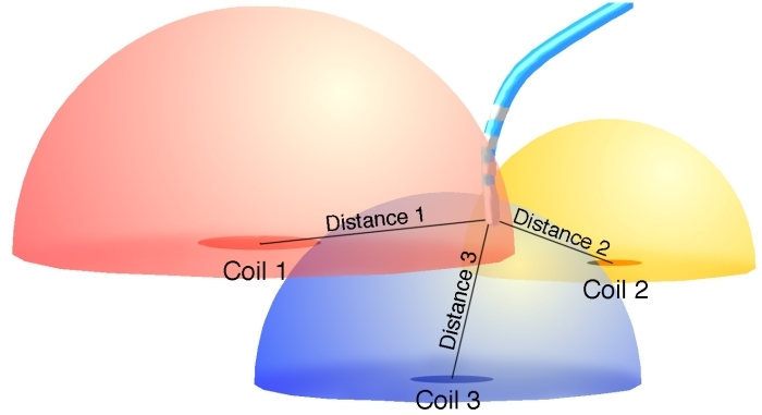
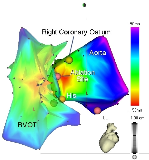
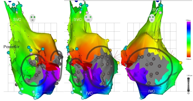
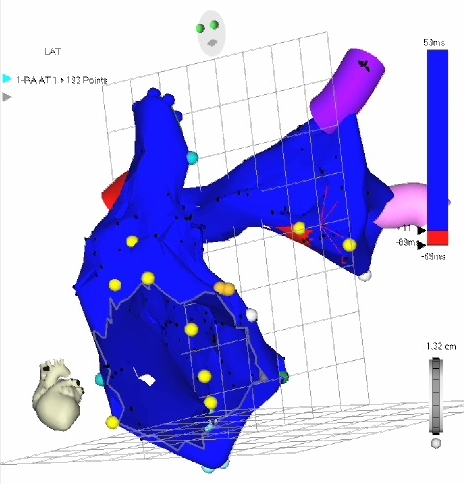
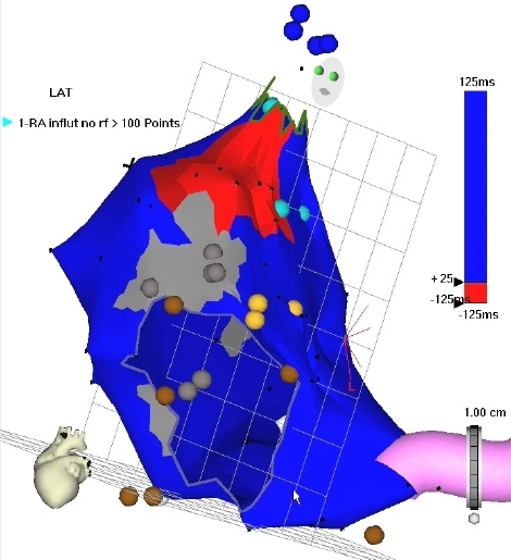
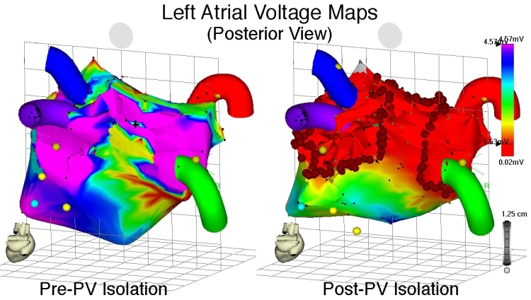
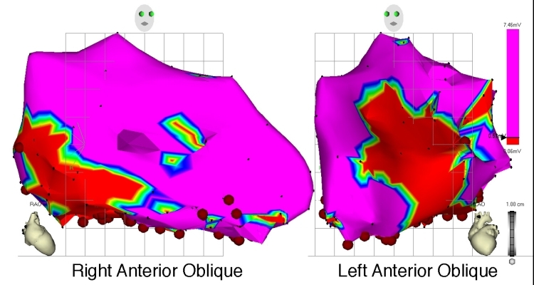
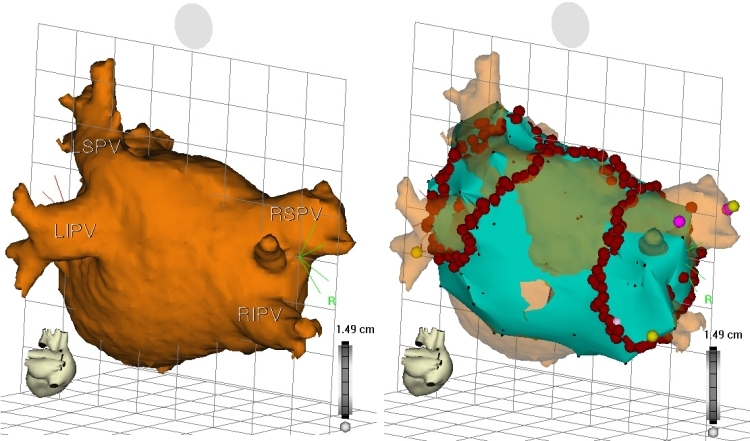
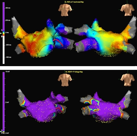
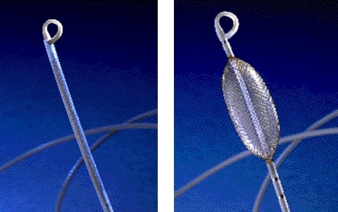
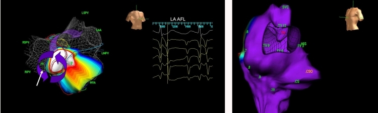
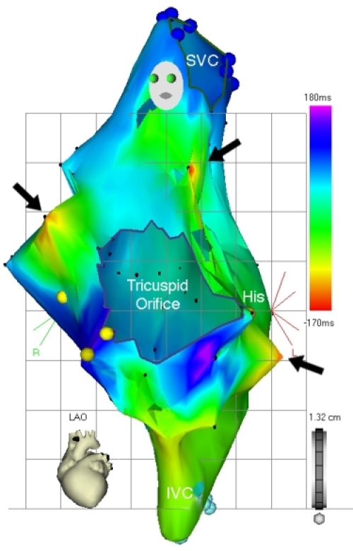
Electrophysiologic testing and radiofrequency ablation have evolved as curative measures for a variety of rhythm disturbances. As experience in this field has grown, ablation is progressively being used to address more complex rhythm disturbances. Paralleling this trend are technological advancements to facilitate these efforts, including electroanatomic mapping (EAM). At present, several different EAM systems utilizing various technologies are available to facilitate mapping and ablation. Use of these systems has been shown to reduce fluoroscopic exposure and radiation dose, with less significant effects on procedural duration and success rates. Among the data provided by EAM are chamber reconstruction, tagging of important anatomic landmarks and ablation lesions, display of diagnostic and mapping catheters without using fluoroscopy, activation mapping, and voltage (or scar) mapping. Several EAM systems have specialized features, such as enhanced ability to map non-sustained or hemodynamically unstable arrhythmias, ability to display diagnostic as well as mapping catheter positions, and wide compatibility with a variety of catheters. Each EAM system has its strengths and weaknesses, and the system chosen must depend upon what data is required for procedural success (activation mapping, substrate mapping, cardiac geometry), the anticipated arrhythmia, the compatibility of the system with adjunctive tools (i.e. diagnostic and ablation catheters), and the operator's familiarity with the selected system. While EAM can offer significant assistance during an EP procedure, their incorrect or inappropriate application can substantially hamper mapping efforts and procedural success, and should not replace careful interpretation of data and strict adherence to electrophysiologic principles.
电生理检查和射频消融已发展成为治疗多种心律失常的手段。随着该领域经验的积累,消融术正逐渐用于处理更复杂的心律失常。与此趋势并行的是有助于这些工作的技术进步,包括电解剖标测(EAM)。目前,有几种利用不同技术的不同EAM系统可用于辅助标测和消融。已证明使用这些系统可减少透视曝光和辐射剂量,对手术时间和成功率的影响较小。EAM提供的数据包括腔室重建、重要解剖标志和消融灶的标记、无需透视即可显示诊断和标测导管、激动标测以及电压(或瘢痕)标测。几种EAM系统具有特殊功能,例如增强标测非持续性或血流动力学不稳定心律失常的能力、显示诊断和标测导管位置的能力以及与各种导管的广泛兼容性。每个EAM系统都有其优缺点,所选系统必须取决于手术成功所需的数据(激动标测、基质标测、心脏几何形状)、预期的心律失常、系统与辅助工具(即诊断和消融导管)的兼容性以及操作者对所选系统的熟悉程度。虽然EAM在电生理手术期间可提供重要帮助,但其不正确或不适当的应用会严重妨碍标测工作和手术成功,且不应取代对数据的仔细解读和对电生理原则的严格遵守。