Li Bin, Lin Michael, Tang Ying, Wang Bing, Wang James H-C
MechanoBiology Laboratory, Department of Orthopaedic Surgery, University of Pittsburgh School of Medicine, 210 Lothrop Street, BST, E1640, Pittsburgh, PA 15213, USA.
J Biomech. 2008 Dec 5;41(16):3349-53. doi: 10.1016/j.jbiomech.2008.09.025. Epub 2008 Nov 12.
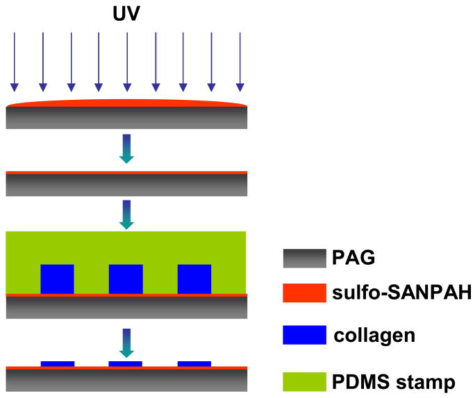
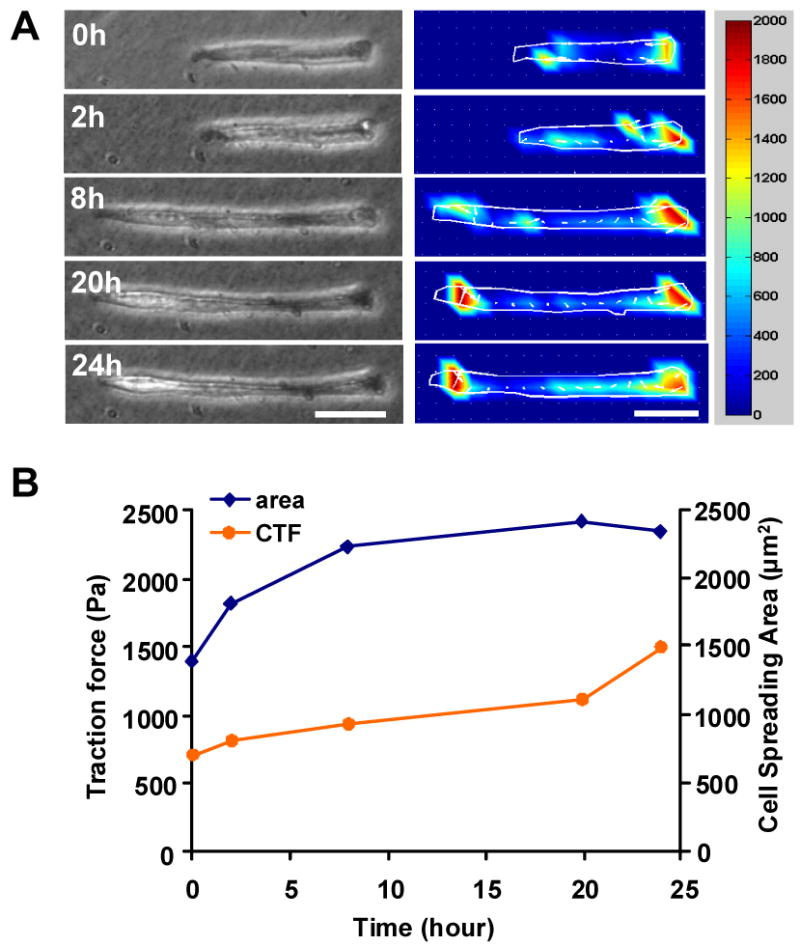
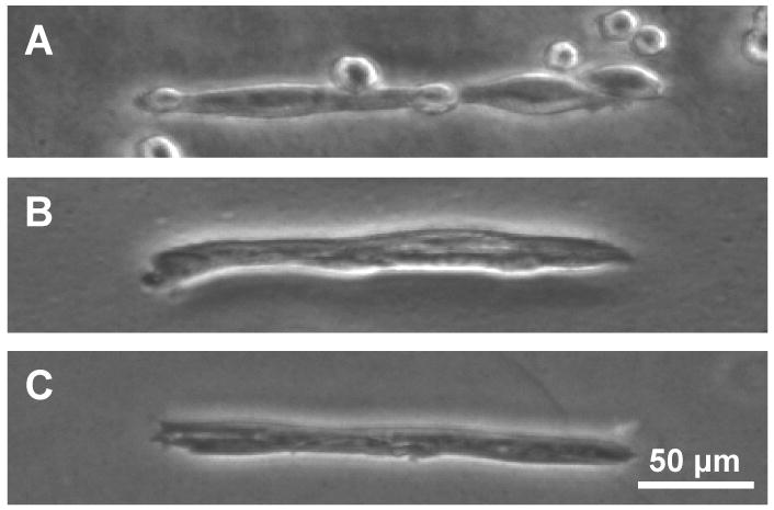
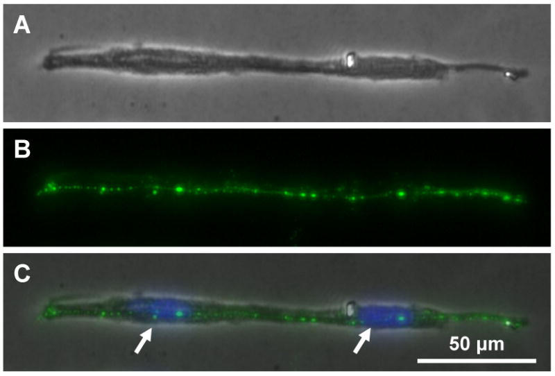
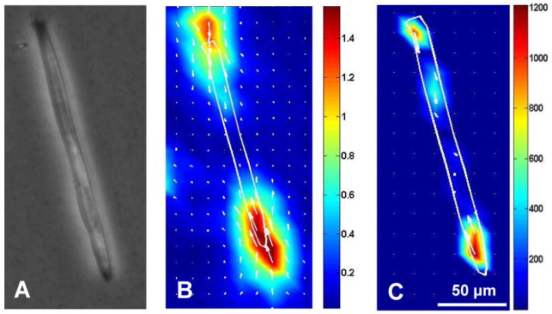
Duchenne muscular dystrophy (DMD) is a lethal disease characterized by rapid, progressive atrophy of muscle tissues. Timely screening of therapeutic interventions is necessary for the development of effective treatment approaches for DMD. We have developed an in vitro model using a combination of micropatterning of C2C12 skeletal muscle cells and cell traction force microscopy (CTFM). In this model, C2C12 cells were micropatterned on a highly elongated adhesive island such that the cells assumed a shape typical of a myotube. During differentiation, these cells gradually fused together and began expressing dystrophin, a structural protein of myotubes, meanwhile, their contractile forces, represented by cell traction forces, continually increased until the myotubes reached maturation. In addition, the high-degree alignment of cells favored myotube differentiation and dystrophin expression. Since the fundamental structural unit of muscle tissue is myofiber, which is responsible for muscle contraction, such a technology that can directly quantify the contractile forces of the myotube, a precursor of myofiber, may constitute a fast and efficient screening approach for DMD therapies.
杜兴氏肌肉营养不良症(DMD)是一种致命疾病,其特征是肌肉组织迅速进行性萎缩。及时筛选治疗干预措施对于开发有效的DMD治疗方法至关重要。我们利用C2C12骨骼肌细胞的微图案化和细胞牵引力显微镜(CTFM)相结合的方法开发了一种体外模型。在这个模型中,C2C12细胞在高度细长的粘附岛上进行微图案化,使细胞呈现出肌管典型的形状。在分化过程中,这些细胞逐渐融合在一起,并开始表达肌营养不良蛋白,这是一种肌管的结构蛋白,与此同时,它们以细胞牵引力表示的收缩力持续增加,直到肌管成熟。此外,细胞的高度排列有利于肌管分化和肌营养不良蛋白表达。由于肌肉组织的基本结构单位是肌纤维,它负责肌肉收缩,这样一种能够直接量化肌纤维前体肌管收缩力的技术,可能构成一种快速有效的DMD治疗筛选方法。