• 文献检索
  • 文档翻译
  • 深度研究
  • 学术资讯
  • Suppr Zotero 插件Zotero 插件
  • 邀请有礼
  • 套餐&价格
  • 历史记录
应用&插件
Suppr Zotero 插件Zotero 插件浏览器插件Mac 客户端Windows 客户端微信小程序
定价
高级版会员购买积分包购买API积分包
服务
文献检索文档翻译深度研究API 文档MCP 服务
关于我们
关于 Suppr公司介绍联系我们用户协议隐私条款
关注我们

Suppr 超能文献

核心技术专利:CN118964589B侵权必究
粤ICP备2023148730 号-1Suppr @ 2026

文献检索

告别复杂PubMed语法,用中文像聊天一样搜索,搜遍4000万医学文献。AI智能推荐,让科研检索更轻松。

立即免费搜索

文件翻译

保留排版,准确专业,支持PDF/Word/PPT等文件格式,支持 12+语言互译。

免费翻译文档

深度研究

AI帮你快速写综述,25分钟生成高质量综述,智能提取关键信息,辅助科研写作。

立即免费体验

利用磁共振图像的自动形态分析对亨廷顿病小鼠模型的脑萎缩进行纵向特征描述。

Longitudinal characterization of brain atrophy of a Huntington's disease mouse model by automated morphological analyses of magnetic resonance images.

机构信息

Department of Radiology, Johns Hopkins University School of Medicine, Baltimore, MD 21205, USA.

出版信息

Neuroimage. 2010 Feb 1;49(3):2340-51. doi: 10.1016/j.neuroimage.2009.10.027. Epub 2009 Oct 19.

DOI:10.1016/j.neuroimage.2009.10.027
PMID:19850133
原文链接:https://pmc.ncbi.nlm.nih.gov/articles/PMC2929697/
Abstract

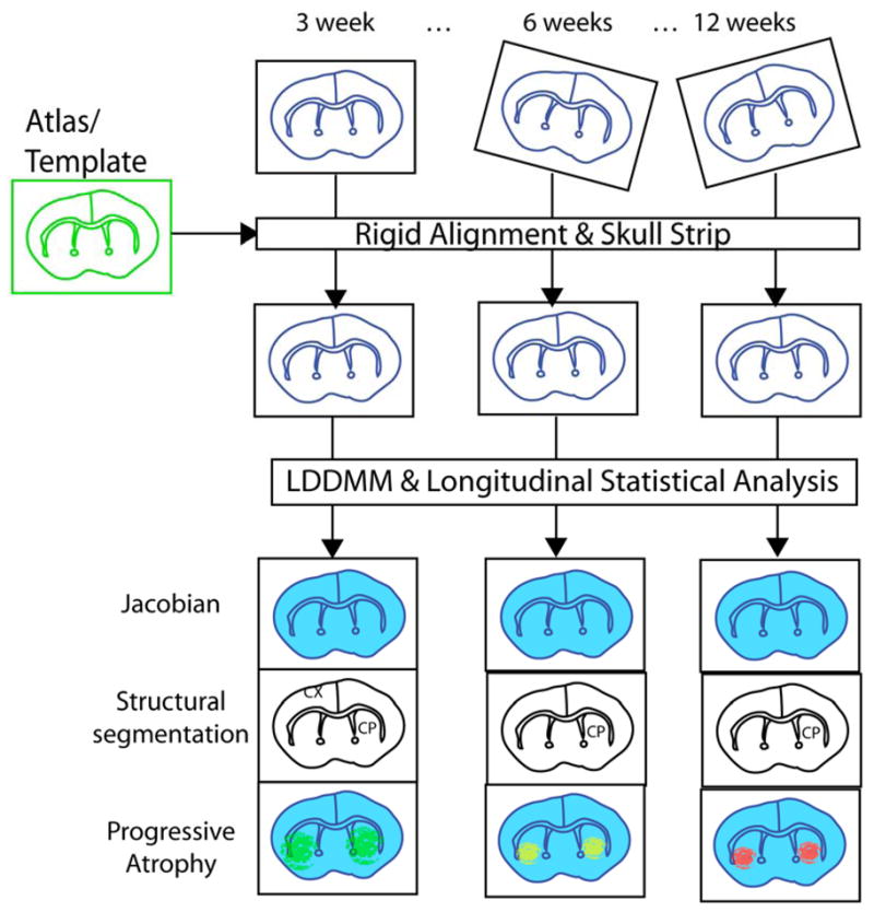
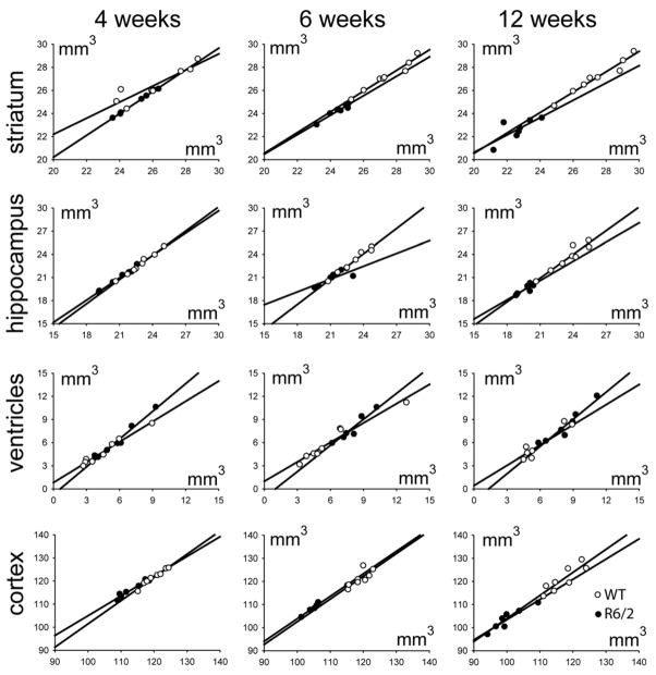
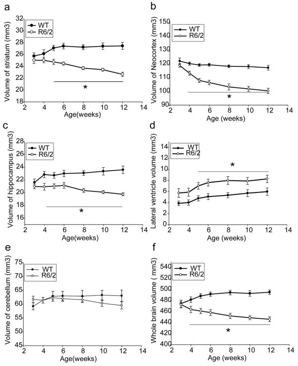
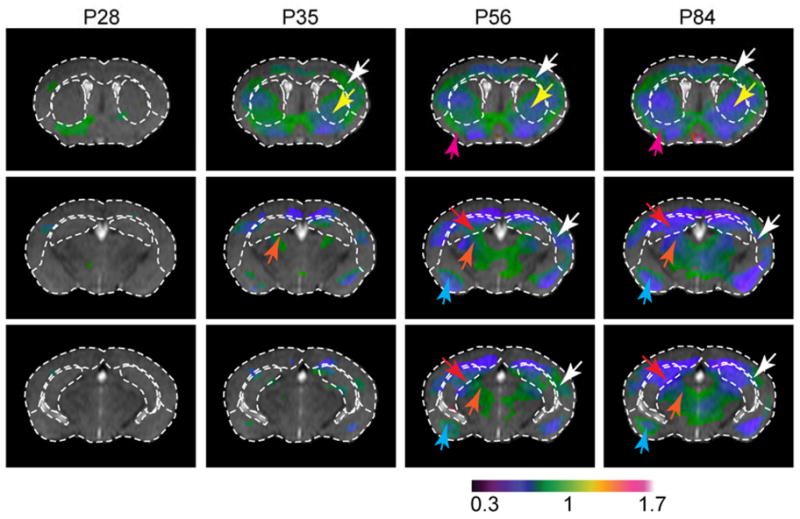
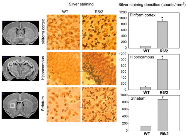
Mouse models of human diseases play crucial roles in understanding disease mechanisms and developing therapeutic measures. Huntington's disease (HD) is characterized by striatal atrophy that begins long before the onset of motor symptoms. In symptomatic HD, striatal volumes decline predictably with disease course. Thus, imaging based volumetric measures have been proposed as outcomes for presymptomatic as well as symptomatic clinical trials of HD. Magnetic resonance imaging of the mouse brain structures is becoming widely available and has been proposed as one of the biomarkers of disease progression and drug efficacy testing. However, three-dimensional and quantitative morphological analyses of the brains are not straightforward. In this paper, we describe a tool for automated segmentation and voxel-based morphological analyses of the mouse brains. This tool was applied to a well-established mouse model of Huntington's disease, the R6/2 transgenic mouse strain. Comparison between the automated and manual segmentation results showed excellent agreement in most brain regions. The automated method was able to sensitively detect atrophy as early as 4 weeks of age and accurately follow disease progression. Comparison between ex vivo and in vivo MRI suggests that the ex vivo end-point measurement of brain morphology is also a valid approach except for the morphology of the ventricles. This is the first report of longitudinal characterization of brain atrophy in a mouse model of Huntington's disease by using automatic morphological analysis.

摘要

人类疾病的小鼠模型在理解疾病机制和开发治疗措施方面发挥着至关重要的作用。亨廷顿病(HD)的特征是纹状体萎缩,这种萎缩早在运动症状出现之前就开始了。在有症状的 HD 中,纹状体体积随着疾病进程的发展可预测性地下降。因此,基于成像的容积测量已被提议作为 HD 无症状和有症状临床试验的结果。对小鼠大脑结构的磁共振成像越来越普及,并已被提议作为疾病进展和药物疗效测试的生物标志物之一。然而,对大脑的三维和定量形态分析并不简单。在本文中,我们描述了一种用于自动分割和基于体素的小鼠大脑形态分析的工具。该工具应用于一种成熟的亨廷顿病小鼠模型,即 R6/2 转基因小鼠品系。自动分割和手动分割结果的比较表明,在大多数脑区都有极好的一致性。自动方法能够敏感地检测到早在 4 周龄时的萎缩,并准确地跟踪疾病的进展。离体和体内 MRI 的比较表明,除了脑室的形态外,脑形态的离体终点测量也是一种有效的方法。这是首例使用自动形态分析对亨廷顿病小鼠模型进行的脑萎缩纵向特征描述。

https://cdn.ncbi.nlm.nih.gov/pmc/blobs/337c/2929697/04127f75e9ac/nihms218508f8.jpg
https://cdn.ncbi.nlm.nih.gov/pmc/blobs/337c/2929697/f3dd9081b1c2/nihms218508f1.jpg
https://cdn.ncbi.nlm.nih.gov/pmc/blobs/337c/2929697/ce071fc82b3f/nihms218508f2.jpg
https://cdn.ncbi.nlm.nih.gov/pmc/blobs/337c/2929697/89263bef46b8/nihms218508f3.jpg
https://cdn.ncbi.nlm.nih.gov/pmc/blobs/337c/2929697/8069945abfef/nihms218508f4.jpg
https://cdn.ncbi.nlm.nih.gov/pmc/blobs/337c/2929697/61812ec453a2/nihms218508f5.jpg
https://cdn.ncbi.nlm.nih.gov/pmc/blobs/337c/2929697/f3a320d9ebc2/nihms218508f6.jpg
https://cdn.ncbi.nlm.nih.gov/pmc/blobs/337c/2929697/59b682628f9c/nihms218508f7.jpg
https://cdn.ncbi.nlm.nih.gov/pmc/blobs/337c/2929697/04127f75e9ac/nihms218508f8.jpg

相似文献

1
Longitudinal characterization of brain atrophy of a Huntington's disease mouse model by automated morphological analyses of magnetic resonance images.利用磁共振图像的自动形态分析对亨廷顿病小鼠模型的脑萎缩进行纵向特征描述。
Neuroimage. 2010 Feb 1;49(3):2340-51. doi: 10.1016/j.neuroimage.2009.10.027. Epub 2009 Oct 19.
2
Spatiotemporal mapping of brain atrophy in mouse models of Huntington's disease using longitudinal in vivo magnetic resonance imaging.利用纵向活体磁共振成像对亨廷顿病小鼠模型的脑萎缩进行时空映射。
Neuroimage. 2012 May 1;60(4):2086-95. doi: 10.1016/j.neuroimage.2012.01.141. Epub 2012 Feb 9.
3
Structural MRI detects progressive regional brain atrophy and neuroprotective effects in N171-82Q Huntington's disease mouse model.结构磁共振成像检测 N171-82Q 亨廷顿病小鼠模型的进行性区域性脑萎缩和神经保护作用。
Neuroimage. 2011 Jun 1;56(3):1027-34. doi: 10.1016/j.neuroimage.2011.02.022. Epub 2011 Feb 12.
4
Voxel-based morphometry in the R6/2 transgenic mouse reveals differences between genotypes not seen with manual 2D morphometry.基于体素的形态测量学在R6/2转基因小鼠中的应用揭示了基因型之间的差异,而这些差异是二维手动形态测量学所无法发现的。
Neurobiol Dis. 2009 Jan;33(1):20-7. doi: 10.1016/j.nbd.2008.09.016. Epub 2008 Oct 1.
5
Use of magnetic resonance imaging for anatomical phenotyping of the R6/2 mouse model of Huntington's disease.利用磁共振成像对亨廷顿舞蹈病R6/2小鼠模型进行解剖学表型分析。
Neurobiol Dis. 2009 Jan;33(1):12-9. doi: 10.1016/j.nbd.2008.09.017. Epub 2008 Oct 1.
6
Correlations of behavioral deficits with brain pathology assessed through longitudinal MRI and histopathology in the R6/1 mouse model of Huntington's disease.通过纵向 MRI 和组织病理学评估亨廷顿病 R6/1 小鼠模型中的行为缺陷与脑病理学的相关性。
PLoS One. 2013 Dec 19;8(12):e84726. doi: 10.1371/journal.pone.0084726. eCollection 2013.
7
Distribution of grey matter atrophy in Huntington's disease patients: a combined ROI-based and voxel-based morphometric study.亨廷顿舞蹈症患者灰质萎缩的分布:一项基于感兴趣区和体素的形态学联合研究
Neuroimage. 2006 Oct 1;32(4):1562-75. doi: 10.1016/j.neuroimage.2006.05.057. Epub 2006 Jul 27.
8
Accuracy of automated amygdala MRI segmentation approaches in Huntington's disease in the IMAGE-HD cohort.亨廷顿病 IMAGE-HD 队列中基于 MRI 的自动杏仁核分割方法的准确性。
Hum Brain Mapp. 2020 May;41(7):1875-1888. doi: 10.1002/hbm.24918. Epub 2020 Feb 7.
9
An independent study of the preclinical efficacy of C2-8 in the R6/2 transgenic mouse model of Huntington's disease.关于C2-8在亨廷顿舞蹈病R6/2转基因小鼠模型中的临床前疗效的独立研究。
J Huntingtons Dis. 2013;2(4):443-51. doi: 10.3233/JHD-130074.
10
Network topology and functional connectivity disturbances precede the onset of Huntington's disease.网络拓扑结构和功能连接性紊乱先于亨廷顿舞蹈症的发病。
Brain. 2015 Aug;138(Pt 8):2332-46. doi: 10.1093/brain/awv145. Epub 2015 Jun 9.

引用本文的文献

1
Considerations and recommendations from the ISMRM diffusion study group for preclinical diffusion MRI: Part 2-Ex vivo imaging: Added value and acquisition.国际磁共振医学学会(ISMRM)扩散研究小组关于临床前扩散磁共振成像的考量与建议:第2部分——离体成像:附加价值与采集
Magn Reson Med. 2025 Jun;93(6):2535-2560. doi: 10.1002/mrm.30435. Epub 2025 Mar 4.
2
The effects of locus coeruleus ablation on mouse brain volume and microstructure evaluated by high-field MRI.通过高场磁共振成像评估蓝斑核消融对小鼠脑容量和微观结构的影响。
Front Cell Neurosci. 2024 Dec 11;18:1498133. doi: 10.3389/fncel.2024.1498133. eCollection 2024.
3
Magnetic Resonance Imaging to Detect Structural Brain Changes in Huntington's Disease: A Review of Data from Mouse Models.

本文引用的文献

1
Longitudinal MRI monitoring of brain damage in the neonatal ventral hippocampal lesion rat model of schizophrenia.精神分裂症幼鼠腹侧海马损伤模型的纵向 MRI 脑损伤监测。
Hippocampus. 2010 Feb;20(2):264-78. doi: 10.1002/hipo.20628.
2
Atlas-based whole brain white matter analysis using large deformation diffeomorphic metric mapping: application to normal elderly and Alzheimer's disease participants.基于图谱的全脑白质分析,采用大变形微分同胚度量映射:应用于正常老年人和阿尔茨海默病患者。
Neuroimage. 2009 Jun;46(2):486-99. doi: 10.1016/j.neuroimage.2009.01.002.
3
Longitudinal regional brain volume changes quantified in normal aging and Alzheimer's APP x PS1 mice using MRI.
磁共振成像检测亨廷顿病的结构性脑变化:来自小鼠模型的数据综述。
J Huntingtons Dis. 2024;13(3):279-299. doi: 10.3233/JHD-240045.
4
The contribution of preclinical magnetic resonance imaging and spectroscopy to Huntington's disease.临床前磁共振成像和光谱学对亨廷顿舞蹈病的贡献。
Front Aging Neurosci. 2024 Feb 13;16:1306312. doi: 10.3389/fnagi.2024.1306312. eCollection 2024.
5
A new open, high-resolution, multishell, diffusion-weighted imaging dataset of the living squirrel monkey.活体松鼠猴的新型开放式、高分辨率、多壳层、弥散加权成像数据集。
Sci Data. 2023 Apr 20;10(1):224. doi: 10.1038/s41597-023-02126-x.
6
A Novel Huntington's Disease Assessment Platform to Support Future Drug Discovery and Development.一种新型亨廷顿舞蹈症评估平台,旨在支持未来的药物发现和开发。
Int J Mol Sci. 2022 Nov 25;23(23):14763. doi: 10.3390/ijms232314763.
7
Thalamocortical Projections Are Significantly Impaired in the R6/2 Mouse Model of Huntington's Disease.亨廷顿病 R6/2 小鼠模型中丘脑皮质投射明显受损。
eNeuro. 2022 Jun 22;9(3). doi: 10.1523/ENEURO.0103-22.2022. Print 2022 May-Jun.
8
Huntingtin and the Synapse.亨廷顿蛋白与突触
Front Cell Neurosci. 2021 Jun 15;15:689332. doi: 10.3389/fncel.2021.689332. eCollection 2021.
9
Huntingtin silencing delays onset and slows progression of Huntington's disease: a biomarker study.亨廷顿病发病延迟和进展减缓的亨廷顿基因沉默:一项生物标志物研究。
Brain. 2021 Nov 29;144(10):3101-3113. doi: 10.1093/brain/awab190.
10
Neuroimaging, Urinary, and Plasma Biomarkers of Treatment Response in Huntington's Disease: Preclinical Evidence with the p75 Ligand LM11A-31.神经影像学、尿液和血浆生物标志物在亨廷顿病治疗反应中的作用:p75 配体 LM11A-31 的临床前证据。
Neurotherapeutics. 2021 Apr;18(2):1039-1063. doi: 10.1007/s13311-021-01023-8. Epub 2021 Mar 30.
利用磁共振成像(MRI)对正常衰老小鼠和阿尔茨海默病APP x PS1小鼠的脑区纵向体积变化进行量化。
Brain Res. 2009 May 13;1270:19-32. doi: 10.1016/j.brainres.2009.02.045. Epub 2009 Mar 9.
4
Automated segmentation of mouse brain images using extended MRF.使用扩展马尔可夫随机场对小鼠脑图像进行自动分割。
Neuroimage. 2009 Jul 1;46(3):717-25. doi: 10.1016/j.neuroimage.2009.02.012. Epub 2009 Feb 21.
5
Analysis of serial magnetic resonance images of mouse brains using image registration.使用图像配准技术对小鼠大脑的系列磁共振图像进行分析。
Neuroimage. 2009 Feb 1;44(3):692-700. doi: 10.1016/j.neuroimage.2008.10.016. Epub 2008 Oct 29.
6
Voxel-based morphometry in the R6/2 transgenic mouse reveals differences between genotypes not seen with manual 2D morphometry.基于体素的形态测量学在R6/2转基因小鼠中的应用揭示了基因型之间的差异,而这些差异是二维手动形态测量学所无法发现的。
Neurobiol Dis. 2009 Jan;33(1):20-7. doi: 10.1016/j.nbd.2008.09.016. Epub 2008 Oct 1.
7
Use of magnetic resonance imaging for anatomical phenotyping of the R6/2 mouse model of Huntington's disease.利用磁共振成像对亨廷顿舞蹈病R6/2小鼠模型进行解剖学表型分析。
Neurobiol Dis. 2009 Jan;33(1):12-9. doi: 10.1016/j.nbd.2008.09.017. Epub 2008 Oct 1.
8
Differential MRI patterns of brain atrophy in double or single transgenic mice for APP and/or SOD.APP和/或SOD双转基因或单转基因小鼠脑萎缩的磁共振成像差异模式
J Neurosci Res. 2008 Nov 15;86(15):3275-84. doi: 10.1002/jnr.21778.
9
Longitudinal brain MRI study in a mouse model of Rett Syndrome and the effects of choline.雷特综合征小鼠模型的纵向脑磁共振成像研究及胆碱的作用。
Neurobiol Dis. 2008 Jul;31(1):110-9. doi: 10.1016/j.nbd.2008.03.009. Epub 2008 Apr 10.
10
Longitudinal neuroanatomical changes determined by deformation-based morphometry in a mouse model of Alzheimer's disease.在阿尔茨海默病小鼠模型中,通过基于变形的形态测量法确定的纵向神经解剖学变化。
Neuroimage. 2008 Aug 1;42(1):19-27. doi: 10.1016/j.neuroimage.2008.04.252. Epub 2008 May 7.