Joo Chulmin, Evans Conor L, Stepinac Thomas, Hasan Tayyaba, de Boer Johannes F
Wellman Center for Photomedicine, Harvard Medical School and Massachusetts General Hospital, Boston, Massachusetts 02114, USA.
Opt Express. 2010 Feb 1;18(3):2858-71. doi: 10.1364/OE.18.002858.
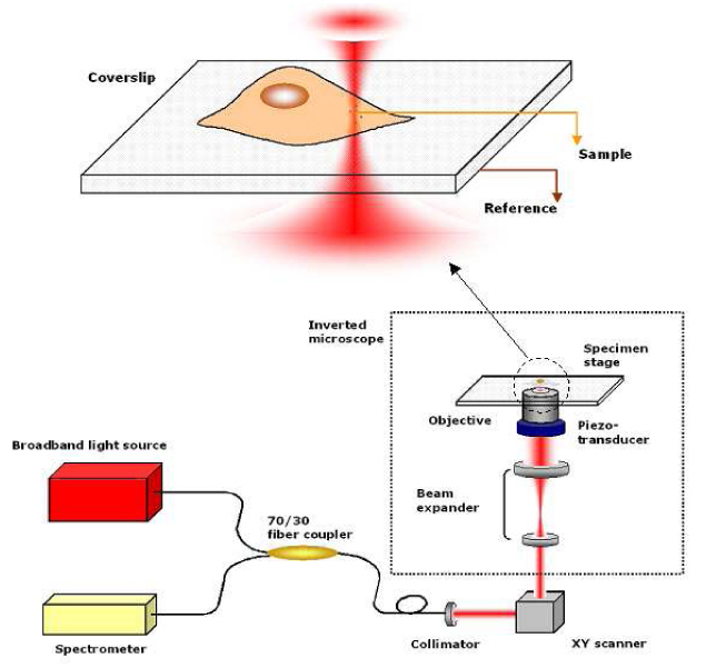
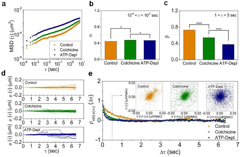
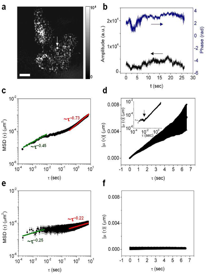
Quantitative measurement of diffusive and directional processes of intracellular structures is not only critical in understanding cell mechanics and functions, but also has many applications, such as investigation of cellular responses to therapeutic agents. We introduce a label-free optical technique that allows non-perturbative characterization of localized intracellular dynamics. The method combines a field-based dynamic light scattering analysis with a confocal interferometric microscope to provide a statistical measure of the diffusive and directional motion of scattering structures inside a microscopic probe volume. To demonstrate the potential of this technique, we examined the localized intracellular dynamics in human epithelial ovarian cancer cells. We observed the distinctive temporal regimes of intracellular dynamics, which transitions from random to directional processes on a timescale of ~0.01 sec. In addition, we observed disrupted directional processes on the timescale of 1 approximately 5 sec by the application of a microtubule polymerization inhibitor, Colchicine, and ATP depletion.
对细胞内结构的扩散和定向过程进行定量测量不仅对于理解细胞力学和功能至关重要,而且还有许多应用,例如研究细胞对治疗药物的反应。我们介绍一种无标记光学技术,该技术能够对局部细胞内动力学进行非侵入性表征。该方法将基于场的动态光散射分析与共聚焦干涉显微镜相结合,以提供对微观探测体积内散射结构的扩散和定向运动的统计测量。为了证明该技术的潜力,我们研究了人上皮性卵巢癌细胞中的局部细胞内动力学。我们观察到细胞内动力学的独特时间模式,其在约0.01秒的时间尺度上从随机过程转变为定向过程。此外,通过应用微管聚合抑制剂秋水仙碱和ATP耗竭,我们在1至5秒的时间尺度上观察到定向过程受到破坏。