Institute of Information Science, Academia Sinica, Taipei, Taiwan.
Bioinformatics. 2010 Jun 15;26(12):i29-37. doi: 10.1093/bioinformatics/btq194.
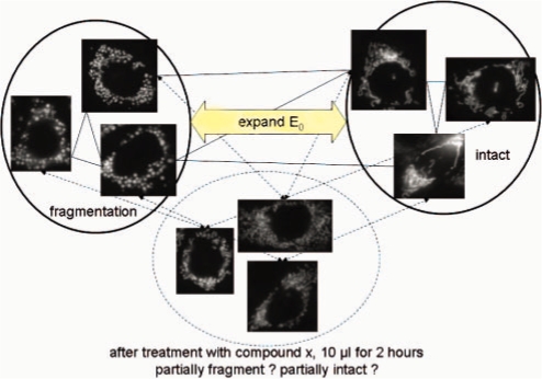
High-throughput image-based assay technologies can rapidly produce a large number of cell images for drug screening, but data analysis is still a major bottleneck that limits their utility. Quantifying a wide variety of morphological differences observed in cell images under different drug influences is still a challenging task because the result can be highly sensitive to sampling and noise.
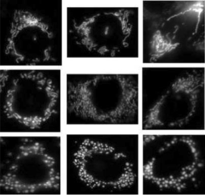
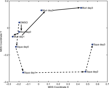
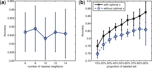
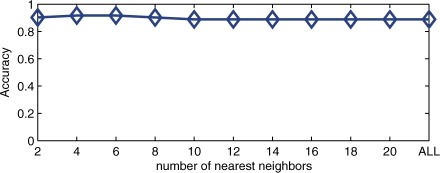
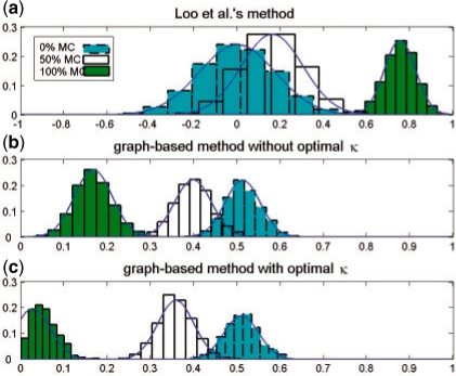
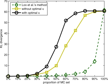
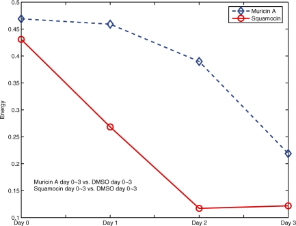
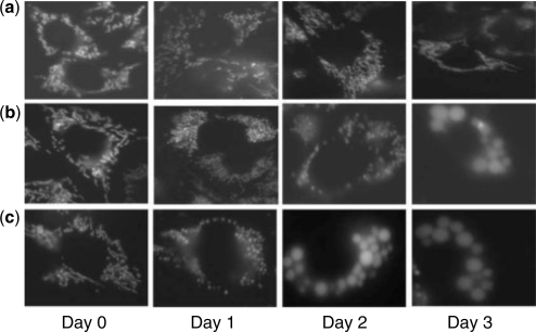
We propose a graph-based approach to cell image analysis. We define graph transition energy to quantify morphological differences between image sets. A spectral graph theoretic regularization is applied to transform the feature space based on training examples of extremely different images to calibrate the quantification. Calibration is essential for a practical quantification method because we need to measure the confidence of the quantification. We applied our method to quantify the degree of partial fragmentation of mitochondria in collections of fluorescent cell images. We show that with transformation, the quantification can be more accurate and sensitive than that without transformation. We also show that our method outperforms competing methods, including neighbourhood component analysis and the multi-variate drug profiling method by Loo et al. We illustrate its utility with a study of Annonaceous acetogenins, a family of compounds with drug potential. Our result reveals that squamocin induces more fragmented mitochondria than muricin A.
Mitochondrial cell images, their corresponding feature sets (SSLF and WSLF) and the source code of our proposed method are available at http://aiia.iis.sinica.edu.tw/.
Supplementary data are available at Bioinformatics online.
高通量基于图像的测定技术可以快速生成大量用于药物筛选的细胞图像,但数据分析仍然是限制其应用的主要瓶颈。由于结果对采样和噪声高度敏感,因此量化不同药物影响下细胞图像中观察到的各种形态差异仍然是一项具有挑战性的任务。
我们提出了一种基于图的细胞图像分析方法。我们定义图转换能量来量化图像集之间的形态差异。应用谱图理论正则化将基于非常不同图像的训练示例的特征空间转换来校准定量。校准对于实用的定量方法至关重要,因为我们需要测量定量的置信度。我们将我们的方法应用于量化荧光细胞图像集合中线粒体的部分碎片化程度。我们表明,通过转换,定量可以比没有转换更准确和敏感。我们还表明,我们的方法优于竞争方法,包括邻域成分分析和 Loo 等人的多变量药物分析方法。我们通过对 Annonaceous 乙酰基化合物(具有药物潜力的化合物家族)的研究说明了其用途。我们的结果表明, squamocin 诱导的线粒体碎片化程度高于 muricin A。
线粒体细胞图像、它们对应的特征集(SSLF 和 WSLF)以及我们提出的方法的源代码可在 http://aiia.iis.sinica.edu.tw/ 获得。
补充数据可在生物信息学在线获得。