NanoScience Technology Center, University of Central Florida, Orlando, Florida, USA.
PLoS One. 2010 Jun 10;5(6):e11042. doi: 10.1371/journal.pone.0011042.
To date, biological components have been incorporated into MEMS devices to create cell-based sensors and assays, motors and actuators, and pumps. Bio-MEMS technologies present a unique opportunity to study fundamental biological processes at a level unrealized with previous methods. The capability to miniaturize analytical systems enables researchers to perform multiple experiments in parallel and with a high degree of control over experimental variables for high-content screening applications.
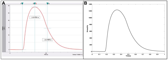
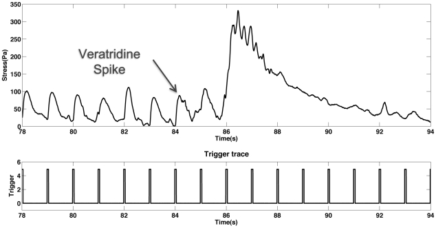
METHODOLOGY/PRINCIPAL FINDINGS: We have demonstrated a biological microelectromechanical system (BioMEMS) based on silicon cantilevers and an AFM detection system for studying the physiology and kinetics of myotubes derived from embryonic rat skeletal muscle. It was shown that it is possible to interrogate and observe muscle behavior in real time, as well as selectively stimulate the contraction of myotubes with the device. Stress generation of the tissue was estimated using a modification of Stoney's equation. Calculated stress values were in excellent agreement with previously published results for cultured myotubes, but not adult skeletal muscle. Other parameters such as time to peak tension (TPT), the time to half relaxation ((1/2)RT) were compared to the literature. It was observed that the myotubes grown on the BioMEMS device, while generating stress magnitudes comparable to those previously published, exhibited slower TPT and (1/2)RT values. However, growth in an enhanced media increased these values. From these data it was concluded that the myotubes cultured on the cantilevers were of an embryonic phenotype. The system was also shown to be responsive to the application of a toxin, veratridine.
CONCLUSIONS/SIGNIFICANCE: The device demonstrated here will provide a useful foundation for studying various aspects of muscle physiology and behavior in a controlled high-throughput manner as well as be useful for biosensor and drug discovery applications.
迄今为止,生物成分已被整合到 MEMS 器件中,以创建基于细胞的传感器和分析、电机和致动器以及泵。生物 MEMS 技术为在以前的方法无法实现的水平上研究基本生物学过程提供了独特的机会。将分析系统微型化的能力使研究人员能够并行进行多项实验,并对实验变量进行高度控制,以实现高通量筛选应用。
方法/主要发现:我们展示了一种基于硅悬臂梁和原子力显微镜检测系统的生物微机电系统 (BioMEMS),用于研究源自胚胎大鼠骨骼肌的肌管的生理学和动力学。结果表明,有可能实时询问和观察肌肉行为,并且可以使用该设备选择性地刺激肌管收缩。使用 Stoney 方程的修改来估计组织的应力产生。计算出的应力值与先前发表的培养肌管的结果非常吻合,但与成年骨骼肌不符。还将其他参数(如峰值张力时间 (TPT)、半松弛时间 ((1/2)RT) 与文献进行了比较。观察到在 BioMEMS 设备上生长的肌管虽然产生的应力值与以前发表的结果相当,但 TPT 和 (1/2)RT 值较慢。然而,在增强的培养基中生长会增加这些值。从这些数据中得出结论,在悬臂上培养的肌管具有胚胎表型。该系统还被证明对毒素藜芦碱的应用有反应。
结论/意义:这里展示的设备将为以受控的高通量方式研究肌肉生理学和行为的各个方面提供有用的基础,并且对生物传感器和药物发现应用也很有用。