• 文献检索
  • 文档翻译
  • 深度研究
  • 学术资讯
  • Suppr Zotero 插件Zotero 插件
  • 邀请有礼
  • 套餐&价格
  • 历史记录
应用&插件
Suppr Zotero 插件Zotero 插件浏览器插件Mac 客户端Windows 客户端微信小程序
定价
高级版会员购买积分包购买API积分包
服务
文献检索文档翻译深度研究API 文档MCP 服务
关于我们
关于 Suppr公司介绍联系我们用户协议隐私条款
关注我们

Suppr 超能文献

核心技术专利:CN118964589B侵权必究
粤ICP备2023148730 号-1Suppr @ 2026

文献检索

告别复杂PubMed语法,用中文像聊天一样搜索,搜遍4000万医学文献。AI智能推荐,让科研检索更轻松。

立即免费搜索

文件翻译

保留排版,准确专业,支持PDF/Word/PPT等文件格式,支持 12+语言互译。

免费翻译文档

深度研究

AI帮你快速写综述,25分钟生成高质量综述,智能提取关键信息,辅助科研写作。

立即免费体验

用于细胞群体力学的亚细胞黏弹性模型。

A sub-cellular viscoelastic model for cell population mechanics.

机构信息

Molecular Cell Biomechanics Laboratory, Department of Bioengineering, University of California, Berkeley, California, United States of America.

出版信息

PLoS One. 2010 Aug 10;5(8):e12097. doi: 10.1371/journal.pone.0012097.

DOI:10.1371/journal.pone.0012097
PMID:20856895
原文链接:https://pmc.ncbi.nlm.nih.gov/articles/PMC2938372/
Abstract

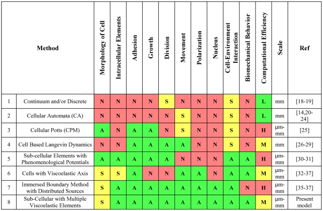
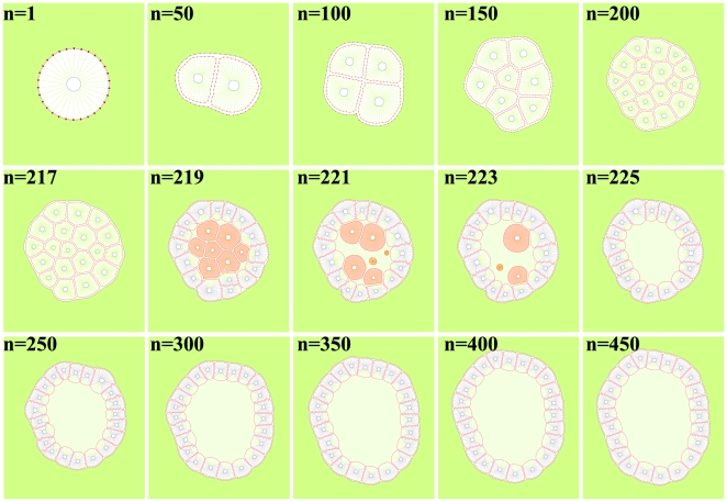
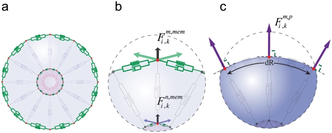
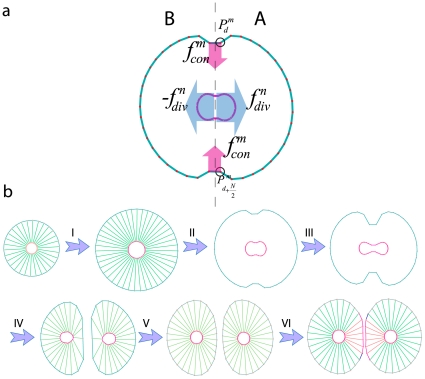
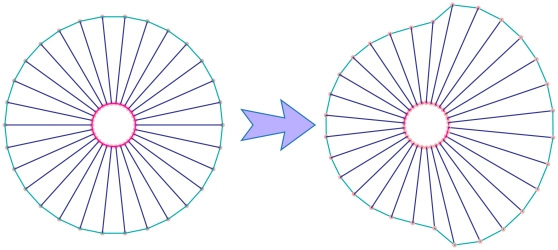
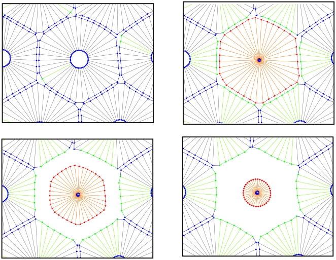
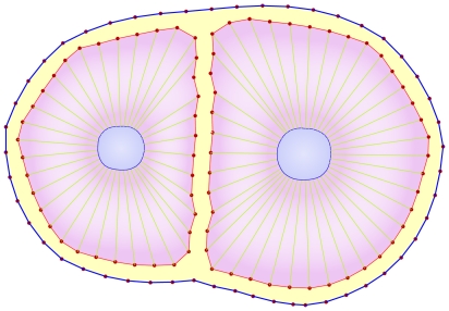
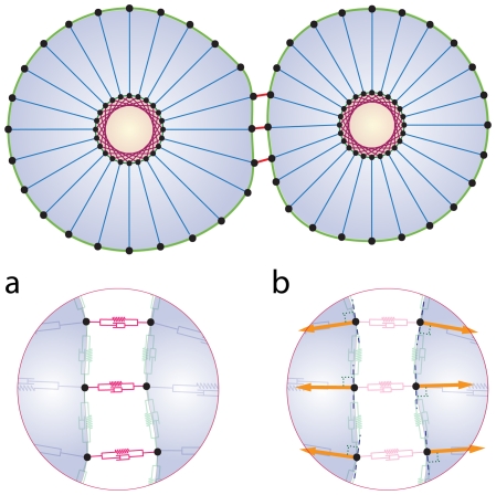
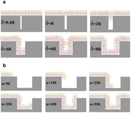
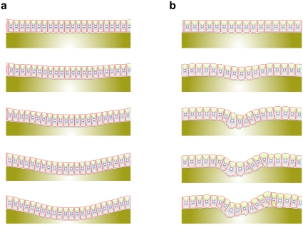
Understanding the biomechanical properties and the effect of biomechanical force on epithelial cells is key to understanding how epithelial cells form uniquely shaped structures in two or three-dimensional space. Nevertheless, with the limitations and challenges posed by biological experiments at this scale, it becomes advantageous to use mathematical and 'in silico' (computational) models as an alternate solution. This paper introduces a single-cell-based model representing the cross section of a typical tissue. Each cell in this model is an individual unit containing several sub-cellular elements, such as the elastic plasma membrane, enclosed viscoelastic elements that play the role of cytoskeleton, and the viscoelastic elements of the cell nucleus. The cell membrane is divided into segments where each segment (or point) incorporates the cell's interaction and communication with other cells and its environment. The model is capable of simulating how cells cooperate and contribute to the overall structure and function of a particular tissue; it mimics many aspects of cellular behavior such as cell growth, division, apoptosis and polarization. The model allows for investigation of the biomechanical properties of cells, cell-cell interactions, effect of environment on cellular clusters, and how individual cells work together and contribute to the structure and function of a particular tissue. To evaluate the current approach in modeling different topologies of growing tissues in distinct biochemical conditions of the surrounding media, we model several key cellular phenomena, namely monolayer cell culture, effects of adhesion intensity, growth of epithelial cell through interaction with extra-cellular matrix (ECM), effects of a gap in the ECM, tensegrity and tissue morphogenesis and formation of hollow epithelial acini. The proposed computational model enables one to isolate the effects of biomechanical properties of individual cells and the communication between cells and their microenvironment while simultaneously allowing for the formation of clusters or sheets of cells that act together as one complex tissue.

摘要

了解生物力学特性以及生物力学对上皮细胞的影响对于理解上皮细胞如何在二维或三维空间中形成独特形状的结构至关重要。然而,由于在这种规模的生物学实验中存在限制和挑战,使用数学和“计算机模拟”(计算)模型作为替代解决方案变得具有优势。本文引入了一个基于单细胞的模型,代表了典型组织的横截面。该模型中的每个细胞都是一个包含几个亚细胞元件的独立单元,例如弹性质膜、起细胞骨架作用的粘性弹性元件以及细胞核的粘性弹性元件。细胞膜被分为若干段,每段(或点)都包含细胞与其他细胞及其环境的相互作用和通讯。该模型能够模拟细胞如何合作并为特定组织的整体结构和功能做出贡献;它模拟了细胞行为的许多方面,例如细胞生长、分裂、凋亡和极化。该模型允许研究细胞的生物力学特性、细胞-细胞相互作用、环境对细胞簇的影响,以及单个细胞如何协同工作并为特定组织的结构和功能做出贡献。为了评估当前在建模不同拓扑结构的生长组织的方法,我们模拟了几种关键的细胞现象,即单层细胞培养、附着强度的影响、通过与细胞外基质(ECM)相互作用生长的上皮细胞、ECM 中的间隙的影响、张力整合和组织形态发生以及中空上皮腺泡的形成。所提出的计算模型能够分离单个细胞的生物力学特性以及细胞与其微环境之间的通讯的影响,同时允许形成作为一个复杂组织一起作用的细胞簇或薄片。

https://cdn.ncbi.nlm.nih.gov/pmc/blobs/d91b/2938372/0da1a923a6b3/pone.0012097.g015.jpg
https://cdn.ncbi.nlm.nih.gov/pmc/blobs/d91b/2938372/d2ad2e154bfe/pone.0012097.g001.jpg
https://cdn.ncbi.nlm.nih.gov/pmc/blobs/d91b/2938372/aac37b5fa3ea/pone.0012097.g002.jpg
https://cdn.ncbi.nlm.nih.gov/pmc/blobs/d91b/2938372/a1872f4df3e8/pone.0012097.g003.jpg
https://cdn.ncbi.nlm.nih.gov/pmc/blobs/d91b/2938372/ad3369e7dc9b/pone.0012097.g004.jpg
https://cdn.ncbi.nlm.nih.gov/pmc/blobs/d91b/2938372/1b4589cb1aea/pone.0012097.g005.jpg
https://cdn.ncbi.nlm.nih.gov/pmc/blobs/d91b/2938372/0c0cc92c0677/pone.0012097.g006.jpg
https://cdn.ncbi.nlm.nih.gov/pmc/blobs/d91b/2938372/672425858a07/pone.0012097.g007.jpg
https://cdn.ncbi.nlm.nih.gov/pmc/blobs/d91b/2938372/eb51d81db9d9/pone.0012097.g008.jpg
https://cdn.ncbi.nlm.nih.gov/pmc/blobs/d91b/2938372/25eff08692d3/pone.0012097.g009.jpg
https://cdn.ncbi.nlm.nih.gov/pmc/blobs/d91b/2938372/bf5daa140ceb/pone.0012097.g010.jpg
https://cdn.ncbi.nlm.nih.gov/pmc/blobs/d91b/2938372/d5e93eb7d555/pone.0012097.g011.jpg
https://cdn.ncbi.nlm.nih.gov/pmc/blobs/d91b/2938372/e578f009feab/pone.0012097.g012.jpg
https://cdn.ncbi.nlm.nih.gov/pmc/blobs/d91b/2938372/435b3ceb46fd/pone.0012097.g013.jpg
https://cdn.ncbi.nlm.nih.gov/pmc/blobs/d91b/2938372/450b969ea636/pone.0012097.g014.jpg
https://cdn.ncbi.nlm.nih.gov/pmc/blobs/d91b/2938372/0da1a923a6b3/pone.0012097.g015.jpg

相似文献

1
A sub-cellular viscoelastic model for cell population mechanics.用于细胞群体力学的亚细胞黏弹性模型。
PLoS One. 2010 Aug 10;5(8):e12097. doi: 10.1371/journal.pone.0012097.
2
An immersed boundary framework for modelling the growth of individual cells: an application to the early tumour development.用于模拟单个细胞生长的浸入边界框架:在肿瘤早期发展中的应用。
J Theor Biol. 2007 Jul 7;247(1):186-204. doi: 10.1016/j.jtbi.2007.02.019. Epub 2007 Mar 12.
3
The matrix environmental and cell mechanical properties regulate cell migration and contribute to the invasive phenotype of cancer cells.基质的环境和细胞力学特性调节细胞迁移,并有助于癌细胞的侵袭表型。
Rep Prog Phys. 2019 Jun;82(6):064602. doi: 10.1088/1361-6633/ab1628. Epub 2019 Apr 4.
4
Adapting a Plant Tissue Model to Animal Development: Introducing Cell Sliding into VirtualLeaf.将植物组织模型应用于动物发育:引入细胞滑动至 VirtualLeaf。
Bull Math Biol. 2019 Aug;81(8):3322-3341. doi: 10.1007/s11538-019-00599-9. Epub 2019 Mar 29.
5
Probing eukaryotic cell mechanics via mesoscopic simulations.通过介观模拟探究真核细胞力学
PLoS Comput Biol. 2017 Sep 18;13(9):e1005726. doi: 10.1371/journal.pcbi.1005726. eCollection 2017 Sep.
6
Viscoelastic retraction of single living stress fibers and its impact on cell shape, cytoskeletal organization, and extracellular matrix mechanics.单个活应力纤维的粘弹性回缩及其对细胞形状、细胞骨架组织和细胞外基质力学的影响。
Biophys J. 2006 May 15;90(10):3762-73. doi: 10.1529/biophysj.105.071506. Epub 2006 Feb 24.
7
Macromolecular crowding: chemistry and physics meet biology (Ascona, Switzerland, 10-14 June 2012).大分子拥挤现象:化学与物理邂逅生物学(瑞士阿斯科纳,2012年6月10日至14日)
Phys Biol. 2013 Aug;10(4):040301. doi: 10.1088/1478-3975/10/4/040301. Epub 2013 Aug 2.
8
Vertex dynamics simulations of viscosity-dependent deformation during tissue morphogenesis.组织形态发生过程中粘度相关变形的顶点动力学模拟。
Biomech Model Mechanobiol. 2015 Apr;14(2):413-25. doi: 10.1007/s10237-014-0613-5. Epub 2014 Sep 17.
9
A quantitative high-resolution computational mechanics cell model for growing and regenerating tissues.用于生长和再生组织的定量高分辨率计算力学细胞模型。
Biomech Model Mechanobiol. 2020 Feb;19(1):189-220. doi: 10.1007/s10237-019-01204-7. Epub 2019 Nov 20.
10
From cells to tissue: A continuum model of epithelial mechanics.从细胞到组织:上皮力学的连续统模型。
Phys Rev E. 2017 Aug;96(2-1):022418. doi: 10.1103/PhysRevE.96.022418. Epub 2017 Aug 31.

引用本文的文献

1
An Insight into Cancer Cells and Disease Progression Through the Lens of Mathematical Modeling.透过数学建模视角洞察癌细胞与疾病进展
Curr Issues Mol Biol. 2025 Jun 20;47(7):477. doi: 10.3390/cimb47070477.
2
Buckling forces and the wavy folds between pleural epithelial cells.板层上皮细胞之间的褶皱和弯曲力。
Biosystems. 2024 Jun;240:105216. doi: 10.1016/j.biosystems.2024.105216. Epub 2024 Apr 29.
3
On modeling the multiscale mechanobiology of soft tissues: Challenges and progress.关于软组织多尺度力学生物学建模:挑战与进展

本文引用的文献

1
Actin complexes in the cell nucleus: new stones in an old field.细胞核中的肌动蛋白复合物:旧领域中的新石头。
Histochem Cell Biol. 2010 Jun;133(6):607-26. doi: 10.1007/s00418-010-0701-2. Epub 2010 May 5.
2
Modeling multicellular systems using subcellular elements.使用亚细胞元件对多细胞系统进行建模。
Math Biosci Eng. 2005 Jul;2(3):613-24. doi: 10.3934/mbe.2005.2.613.
3
Averaged implicit hydrodynamic model of semiflexible filaments.半柔性细丝的平均隐式流体动力学模型。
Biophys Rev (Melville). 2022 Aug 15;3(3):031303. doi: 10.1063/5.0085025. eCollection 2022 Sep.
4
The Effect of Substrate Stiffness on Elastic Force Transmission in the Epithelial Monolayers over Short Timescales.短时间尺度下底物硬度对上皮单层中弹力传递的影响
Cell Mol Bioeng. 2023 Jul 13;16(5-6):475-495. doi: 10.1007/s12195-023-00772-0. eCollection 2023 Dec.
5
Biophysical models of early mammalian embryogenesis.早期哺乳动物胚胎发生的生物物理模型。
Stem Cell Reports. 2023 Jan 10;18(1):26-46. doi: 10.1016/j.stemcr.2022.11.021.
6
Quantitative modeling identifies critical cell mechanics driving bile duct lumen formation.定量建模确定了驱动胆管管腔形成的关键细胞力学。
PLoS Comput Biol. 2022 Feb 18;18(2):e1009653. doi: 10.1371/journal.pcbi.1009653. eCollection 2022 Feb.
7
A novel jamming phase diagram links tumor invasion to non-equilibrium phase separation.一种新颖的干扰相图将肿瘤侵袭与非平衡相分离联系起来。
iScience. 2021 Oct 12;24(11):103252. doi: 10.1016/j.isci.2021.103252. eCollection 2021 Nov 19.
8
Bridging from single to collective cell migration: A review of models and links to experiments.从单细胞迁移到群体细胞迁移的衔接:模型综述及其与实验的联系。
PLoS Comput Biol. 2020 Dec 10;16(12):e1008411. doi: 10.1371/journal.pcbi.1008411. eCollection 2020 Dec.
9
Endothelial Cells Morphology in Response to Combined WSS and Biaxial CS: Introduction of Effective Strain Ratio.内皮细胞对联合壁面剪应力和双轴循环拉伸的形态学响应:有效应变比的引入
Cell Mol Bioeng. 2020 May 12;13(6):647-657. doi: 10.1007/s12195-020-00618-z. eCollection 2020 Dec.
10
Transcellular Model for Neutral and Charged Nanoparticles Across an In Vitro Blood-Brain Barrier.跨体外血脑屏障的中性和带电纳米粒子的细胞间模型。
Cardiovasc Eng Technol. 2020 Dec;11(6):607-620. doi: 10.1007/s13239-020-00496-6. Epub 2020 Oct 28.
Phys Rev E Stat Nonlin Soft Matter Phys. 2010 Mar;81(3 Pt 1):031920. doi: 10.1103/PhysRevE.81.031920. Epub 2010 Mar 26.
4
Dynamics and molecular interactions of linker of nucleoskeleton and cytoskeleton (LINC) complex proteins.核骨架-胞质骨架连接蛋白(LINC)复合物蛋白的动力学和分子相互作用。
J Cell Sci. 2009 Nov 15;122(Pt 22):4099-108. doi: 10.1242/jcs.057075. Epub 2009 Oct 20.
5
Mechanical signaling in networks of motor and cytoskeletal proteins.运动蛋白与细胞骨架蛋白网络中的机械信号传导
Annu Rev Biophys. 2009;38:217-34. doi: 10.1146/annurev.biophys.050708.133732.
6
A Simple 1-D Physical Model for the Crawling Nematode Sperm Cell.一种用于爬行线虫精子细胞的简单一维物理模型。
J Stat Phys. 2003 Mar 1;110(3-6):1169-1189. doi: 10.1023/A:1022153028488.
7
Cell fate regulation by coupling mechanical cycles to biochemical signaling pathways.通过将机械循环与生化信号通路相结合来调控细胞命运
Curr Opin Cell Biol. 2009 Feb;21(1):38-46. doi: 10.1016/j.ceb.2009.01.002. Epub 2009 Feb 11.
8
Mechanotransduction at a distance: mechanically coupling the extracellular matrix with the nucleus.远距离机械转导:将细胞外基质与细胞核进行机械偶联
Nat Rev Mol Cell Biol. 2009 Jan;10(1):75-82. doi: 10.1038/nrm2594.
9
Nuclear shape, mechanics, and mechanotransduction.细胞核形态、力学及力传导
Circ Res. 2008 Jun 6;102(11):1307-18. doi: 10.1161/CIRCRESAHA.108.173989.
10
Modeling cell rheology with the Subcellular Element Model.用亚细胞单元模型模拟细胞流变学。
Phys Biol. 2008 Apr 10;5(1):015002. doi: 10.1088/1478-3975/5/1/015002.