Biomedical Image Computing Group, Department of Radiology and Biomedical Imaging, University of California San Francisco, San Francisco, CA 94143-0628, USA.
Brain Struct Funct. 2011 Jan;215(3-4):255-63. doi: 10.1007/s00429-010-0286-5. Epub 2010 Oct 29.
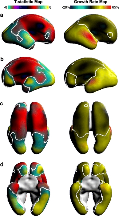
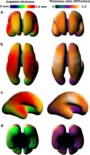
The waiting period of subplate evolution is a critical phase for the proper formation of neural connections in the brain. During this time, which corresponds to 15 to 24 postconceptual weeks (PCW) in the human fetus, thalamocortical and cortico-cortical afferents wait in and are in part guided by molecules embedded in the extracellular matrix of the subplate. Recent advances in fetal MRI techniques now allow us to study the developing brain anatomy in 3D from in utero imaging. We describe a reliable segmentation protocol to delineate the boundaries of the subplate from T2-W MRI. The reliability of the protocol was evaluated in terms of intra-rater reproducibility on a subset of the subjects. We also present the first 3D quantitative analyses of temporal changes in subplate volume, thickness, and contrast from 18 to 24 PCW. Our analysis shows that firstly, global subplate volume increases in proportion with the supratentorial volume; the subplate remained approximately one-third of supratentorial volume. Secondly, we found both global and regional growth in subplate thickness and a linear increase in the median and maximum subplate thickness through the waiting period. Furthermore, we found that posterior regions--specifically the occipital pole, ventral occipito-temporal region, and planum temporale--of the developing brain underwent the most statistically significant increases in subplate thickness. During this period, the thickest region was the developing somatosensory/motor cortex. The subplate growth patterns reported here may be used as a baseline for comparison to abnormal fetal brain development.
基板发育的等待期是大脑中神经连接正确形成的关键阶段。在此期间,即人类胎儿的 15 至 24 孕周(PCW),丘脑皮质和皮质皮质传入纤维在基板的细胞外基质中等待,并部分受嵌入其中的分子的引导。胎儿 MRI 技术的最新进展现在使我们能够从宫内成像中以 3D 形式研究发育中大脑的解剖结构。我们描述了一种可靠的分割协议,用于从 T2-W MRI 中描绘基板的边界。该协议的可靠性是通过对部分受试者的内部评估者可重复性进行评估的。我们还首次对 18 至 24 孕周期间基板体积、厚度和对比度的时间变化进行了 3D 定量分析。我们的分析表明,首先,基板的总体积与颅顶体积成比例增加;基板的体积大约是颅顶体积的三分之一。其次,我们发现基板厚度在整体和局部都在增加,并且在等待期间基板的中位数和最大厚度呈线性增加。此外,我们发现大脑发育过程中后部区域(特别是枕极、腹侧枕颞区和颞平面)的基板厚度增加最为显著。在此期间,最厚的区域是发育中的感觉运动皮层。这里报告的基板生长模式可作为与异常胎儿大脑发育进行比较的基线。