Scott Julia A, Habas Piotr A, Kim Kio, Rajagopalan Vidya, Hamzelou Kia S, Corbett-Detig James M, Barkovich A James, Glenn Orit A, Studholme Colin
Biomedical Image Computing Group, Department of Radiology and Biomedical Imaging, University of California San Francisco, San Francisco, CA, USA.
Int J Dev Neurosci. 2011 Aug;29(5):529-36. doi: 10.1016/j.ijdevneu.2011.04.001. Epub 2011 Apr 17.
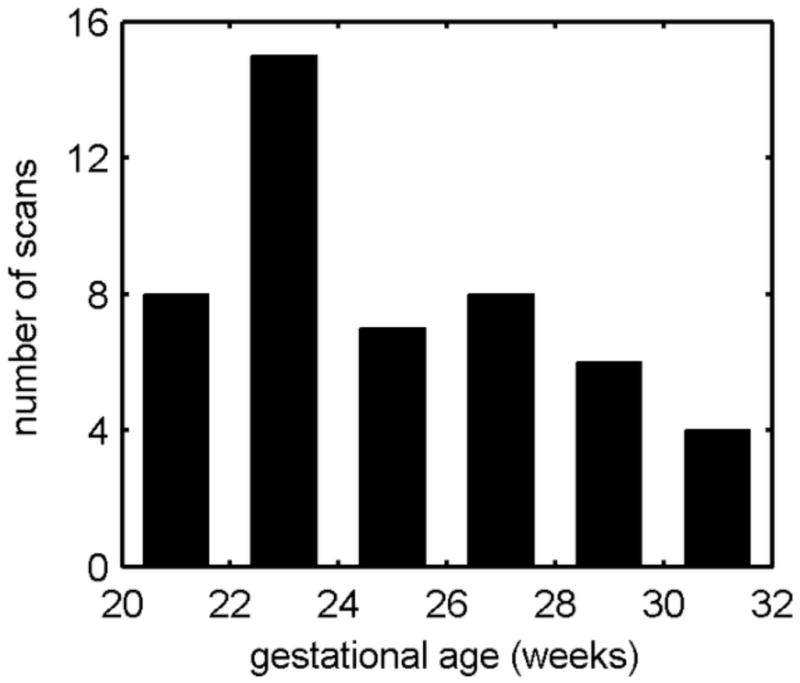
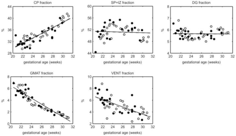
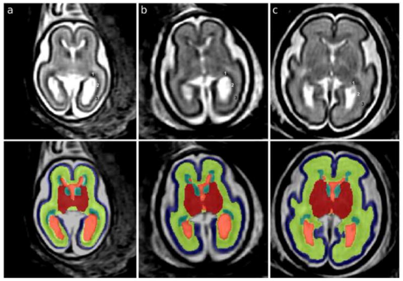
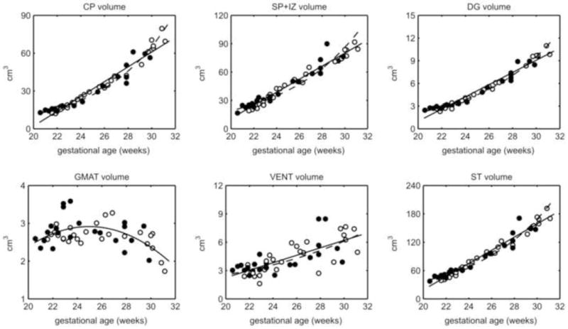
In the latter half of gestation (20-40 gestational weeks), human brain growth accelerates in conjunction with cortical folding and the deceleration of ventricular zone progenitor cell proliferation. These processes are reflected in changes in the volume of respective fetal tissue zones. Thus far, growth trajectories of the fetal tissue zones have been extracted primarily from 2D measurements on histological sections and magnetic resonance imaging (MRI). In this study, the volumes of major fetal zones-cortical plate (CP), subplate and intermediate zone (SP+IZ), germinal matrix (GMAT), deep gray nuclei (DG), and ventricles (VENT)--are calculated from automatic segmentation of motion-corrected, 3D reconstructed MRI. We analyzed 48 T2-weighted MRI scans from 39 normally developing fetuses in utero between 20.57 and 31.14 gestational weeks (GW). The supratentorial volume (STV) increased linearly at a rate of 15.22% per week. The SP+IZ (14.75% per week) and DG (15.56% per week) volumes increased at similar rates. The CP increased at a greater relative rate (18.00% per week), while the VENT (9.18% per week) changed more slowly. Therefore, CP increased as a fraction of STV and the VENT fraction declined. The total GMAT volume slightly increased then decreased after 25 GW. We did not detect volumetric sexual dimorphisms or total hemispheric volume asymmetries, which may emerge later in gestation. Further application of the automated fetal brain segmentation to later gestational ages will bridge the gap between volumetric studies of premature brain development and normal brain development in utero.
在妊娠后半期(妊娠20 - 40周),人类大脑生长加速,同时伴有皮质折叠和脑室区祖细胞增殖减速。这些过程反映在各个胎儿组织区域体积的变化上。迄今为止,胎儿组织区域的生长轨迹主要是从组织学切片和磁共振成像(MRI)的二维测量中提取的。在本研究中,主要胎儿区域——皮质板(CP)、亚板和中间区(SP + IZ)、生发基质(GMAT)、深部灰质核(DG)和脑室(VENT)的体积是通过对运动校正后的三维重建MRI进行自动分割计算得出的。我们分析了39例正常发育胎儿在子宫内妊娠20.57至31.14周(GW)之间的48次T2加权MRI扫描。幕上体积(STV)以每周15.22%的速度线性增加。SP + IZ(每周14.75%)和DG(每周15.56%)的体积以相似的速度增加。CP以更高的相对速度增加(每周18.00%),而VENT(每周9.18%)变化较慢。因此,CP占STV的比例增加,VENT的比例下降。GMAT总体积在25GW后略有增加然后下降。我们未检测到体积上的性别差异或总半球体积不对称,这些可能在妊娠后期出现。将自动胎儿脑分割进一步应用于更大的孕周将弥合早产脑发育和子宫内正常脑发育的体积研究之间的差距。