• 文献检索
  • 文档翻译
  • 深度研究
  • 学术资讯
  • Suppr Zotero 插件Zotero 插件
  • 邀请有礼
  • 套餐&价格
  • 历史记录
应用&插件
Suppr Zotero 插件Zotero 插件浏览器插件Mac 客户端Windows 客户端微信小程序
定价
高级版会员购买积分包购买API积分包
服务
文献检索文档翻译深度研究API 文档MCP 服务
关于我们
关于 Suppr公司介绍联系我们用户协议隐私条款
关注我们

Suppr 超能文献

核心技术专利:CN118964589B侵权必究
粤ICP备2023148730 号-1Suppr @ 2026

文献检索

告别复杂PubMed语法,用中文像聊天一样搜索,搜遍4000万医学文献。AI智能推荐,让科研检索更轻松。

立即免费搜索

文件翻译

保留排版,准确专业,支持PDF/Word/PPT等文件格式,支持 12+语言互译。

免费翻译文档

深度研究

AI帮你快速写综述,25分钟生成高质量综述,智能提取关键信息,辅助科研写作。

立即免费体验

相似文献

1
Computed tomography in color: NanoK-enhanced spectral CT molecular imaging.彩色计算机断层扫描:纳米金增强光谱CT分子成像
Angew Chem Int Ed Engl. 2010 Dec 10;49(50):9635-9. doi: 10.1002/anie.201005657.
2
Dual-modality, fluorescent, PLGA encapsulated bismuth nanoparticles for molecular and cellular fluorescence imaging and computed tomography.用于分子和细胞荧光成像及计算机断层扫描的双模态、荧光、聚乳酸-羟基乙酸共聚物包封的铋纳米颗粒
Nanoscale. 2014 Nov 7;6(21):13104-12. doi: 10.1039/c4nr01405g.
3
Hyaluronic acid-functionalized bismuth oxide nanoparticles for computed tomography imaging-guided radiotherapy of tumor.用于肿瘤计算机断层扫描成像引导放疗的透明质酸功能化氧化铋纳米粒子
Int J Nanomedicine. 2017 Aug 21;12:5973-5992. doi: 10.2147/IJN.S130455. eCollection 2017.
4
Bismuth chelate as a contrast agent for X-ray computed tomography.螯合铋作为 X 射线计算机断层扫描的造影剂。
J Nanobiotechnology. 2020 Aug 6;18(1):110. doi: 10.1186/s12951-020-00669-4.
5
Bismuth drug-inspired ultra-small dextran coated bismuth oxide nanoparticles for targeted computed tomography imaging of inflammatory bowel disease.基于铋药物的超小葡聚糖包覆的氧化铋纳米粒子用于炎症性肠病的靶向计算机断层成像。
Biomaterials. 2024 Dec;311:122658. doi: 10.1016/j.biomaterials.2024.122658. Epub 2024 Jun 7.
6
Ultrasmall Semimetal Nanoparticles of Bismuth for Dual-Modal Computed Tomography/Photoacoustic Imaging and Synergistic Thermoradiotherapy.用于双模式计算机断层扫描/光声成像及协同热放疗的铋的超小半导体纳米粒子。
ACS Nano. 2017 Apr 25;11(4):3990-4001. doi: 10.1021/acsnano.7b00476. Epub 2017 Apr 12.
7
An X-ray computed tomography imaging agent based on long-circulating bismuth sulphide nanoparticles.一种基于长循环硫化铋纳米颗粒的X射线计算机断层扫描成像剂。
Nat Mater. 2006 Feb;5(2):118-22. doi: 10.1038/nmat1571. Epub 2006 Jan 29.
8
Ultrastable polyethyleneimine-stabilized gold nanoparticles modified with polyethylene glycol for blood pool, lymph node and tumor CT imaging.用聚乙二醇修饰的超稳定聚乙烯亚胺稳定金纳米颗粒用于血池、淋巴结和肿瘤的CT成像。
Nanoscale. 2016 Mar 14;8(10):5567-77. doi: 10.1039/c5nr07955a.
9
Harnessing X-Ray Energy-Dependent Attenuation of Bismuth-Based Nanoprobes for Accurate Diagnosis of Liver Fibrosis.利用基于铋的纳米探针的 X 射线能量依赖衰减进行准确的肝纤维化诊断。
Adv Sci (Weinh). 2021 Jun;8(11):e2002548. doi: 10.1002/advs.202002548. Epub 2021 Mar 16.
10
Renal-Clearable Hollow Bismuth Subcarbonate Nanotubes for Tumor Targeted Computed Tomography Imaging and Chemoradiotherapy.用于肿瘤靶向计算机断层扫描成像和放化疗的可肾脏清除的中空碳酸亚铋纳米管。
Nano Lett. 2018 Feb 14;18(2):1196-1204. doi: 10.1021/acs.nanolett.7b04741. Epub 2018 Jan 30.

引用本文的文献

1
Biosensing and Biotechnological Applications of Nanofillers: Current Status and Perspectives.纳米填料的生物传感与生物技术应用:现状与展望
Indian J Microbiol. 2025 Mar;65(1):235-252. doi: 10.1007/s12088-024-01326-2. Epub 2024 Jul 26.
2
Device Design and Advanced Computed Tomography of 3D Printed Radiopaque Composite Scaffolds and Meniscus.3D打印不透射线复合支架和半月板的设备设计与先进计算机断层扫描
Adv Funct Mater. 2024 Oct 29;34(44). doi: 10.1002/adfm.202404860. Epub 2024 May 17.
3
Targeted K-Edge Nanoprobes From Praseodymium and Hafnium for Ratiometric Tracking of Dual Biomarkers using Spectral Photon Counting CT.用于使用光谱光子计数CT对双生物标志物进行比率追踪的镨和铪靶向K边纳米探针。
Adv Sci (Weinh). 2024 Dec;11(46):e2408408. doi: 10.1002/advs.202408408. Epub 2024 Oct 7.
4
Achieving safe and high-performance gastrointestinal tract spectral CT imaging with small-molecule lanthanide complex.利用小分子镧系配合物实现安全、高性能的胃肠道光谱CT成像。
Biomater Res. 2023 Nov 22;27(1):119. doi: 10.1186/s40824-023-00463-x.
5
Clinical Applications of Photon-counting CT: A Review of Pioneer Studies and a Glimpse into the Future.光子计数 CT 的临床应用:先驱研究综述及未来展望。
Radiology. 2023 Oct;309(1):e222432. doi: 10.1148/radiol.222432.
6
Significance of Capping Agents of Colloidal Nanoparticles from the Perspective of Drug and Gene Delivery, Bioimaging, and Biosensing: An Insight.从药物和基因传递、生物成像和生物传感的角度来看胶体纳米粒子封端剂的意义:一种见解。
Int J Mol Sci. 2022 Sep 10;23(18):10521. doi: 10.3390/ijms231810521.
7
Spectral computed tomography with inorganic nanomaterials: State-of-the-art.无机纳米材料的光谱计算机断层扫描:最新技术。
Adv Drug Deliv Rev. 2022 Oct;189:114524. doi: 10.1016/j.addr.2022.114524. Epub 2022 Sep 2.
8
Recent advances in targeted delivery of non-coding RNA-based therapeutics for atherosclerosis.基于非编码 RNA 治疗剂的靶向递呈在动脉粥样硬化中的最新进展。
Mol Ther. 2022 Oct 5;30(10):3118-3132. doi: 10.1016/j.ymthe.2022.07.018. Epub 2022 Aug 1.
9
Fabrication of gelatin BiS capsules as a highly sensitive X-ray contrast agent for gastrointestinal motility assessment .制备明胶双硫胶囊作为用于胃肠动力评估的高灵敏度X射线造影剂。
RSC Adv. 2022 May 5;12(22):13645-13652. doi: 10.1039/d2ra00993e.
10
Prospecting Cellular Gold Nanoparticle Biomineralization as a Viable Alternative to Prefabricated Gold Nanoparticles.探索细胞金纳米颗粒生物矿化作为预制金纳米颗粒的可行替代品。
Adv Sci (Weinh). 2022 Jul;9(20):e2105957. doi: 10.1002/advs.202105957. Epub 2022 May 4.

本文引用的文献

1
Design considerations for tumour-targeted nanoparticles.肿瘤靶向纳米粒子的设计考虑因素。
Nat Nanotechnol. 2010 Jan;5(1):42-7. doi: 10.1038/nnano.2009.314. Epub 2009 Nov 1.
2
Potential of dual-energy computed tomography to characterize atherosclerotic plaque: ex vivo assessment of human coronary arteries in comparison to histology.双能计算机断层扫描在表征动脉粥样硬化斑块方面的潜力:与组织学相比对人体冠状动脉的离体评估。
J Cardiovasc Comput Tomogr. 2008 Jul-Aug;2(4):234-42. doi: 10.1016/j.jcct.2008.05.146. Epub 2008 Jun 12.
3
Experimental feasibility of multi-energy photon-counting K-edge imaging in pre-clinical computed tomography.多能光子计数K边成像在临床前计算机断层扫描中的实验可行性
Phys Med Biol. 2008 Aug 7;53(15):4031-47. doi: 10.1088/0031-9155/53/15/002. Epub 2008 Jul 8.
4
Coronary CT angiography findings in patients without coronary calcification.无冠状动脉钙化患者的冠状动脉CT血管造影结果
AJR Am J Roentgenol. 2008 Jul;191(1):50-5. doi: 10.2214/AJR.07.2954.
5
TiO2 nanoparticles as a soft X-ray molecular probe.二氧化钛纳米颗粒作为一种软X射线分子探针。
Chem Commun (Camb). 2008 Jun 7(21):2471-3. doi: 10.1039/b801392f. Epub 2008 Mar 26.
6
Poor correlation between coronary artery calcification and obstructive coronary artery disease in an end-stage renal disease patient.一名终末期肾病患者的冠状动脉钙化与阻塞性冠状动脉疾病之间的相关性较差。
Hemodial Int. 2008 Jan;12(1):16-22. doi: 10.1111/j.1542-4758.2008.00234.x.
7
Soft and intermediate plaques in coronary arteries: how accurately can we measure CT attenuation using 64-MDCT?冠状动脉中的软斑块和中等密度斑块:使用64层螺旋CT我们能多准确地测量CT衰减值?
AJR Am J Roentgenol. 2007 Oct;189(4):981-8. doi: 10.2214/AJR.07.2296.
8
K-edge imaging in x-ray computed tomography using multi-bin photon counting detectors.使用多通道光子计数探测器的X射线计算机断层扫描中的K边成像。
Phys Med Biol. 2007 Aug 7;52(15):4679-96. doi: 10.1088/0031-9155/52/15/020. Epub 2007 Jul 17.
9
Noninvasive detection of macrophages using a nanoparticulate contrast agent for computed tomography.使用纳米颗粒造影剂进行计算机断层扫描对巨噬细胞的无创检测。
Nat Med. 2007 May;13(5):636-41. doi: 10.1038/nm1571. Epub 2007 Apr 8.
10
Computed tomography calcium quantification as a measure of atherosclerotic plaque morphology and stability.计算机断层扫描钙定量作为动脉粥样硬化斑块形态和稳定性的一种测量方法。
Invest Radiol. 2006 Sep;41(9):674-80. doi: 10.1097/01.rli.0000233325.42572.08.

Computed tomography in color: NanoK-enhanced spectral CT molecular imaging.

作者信息

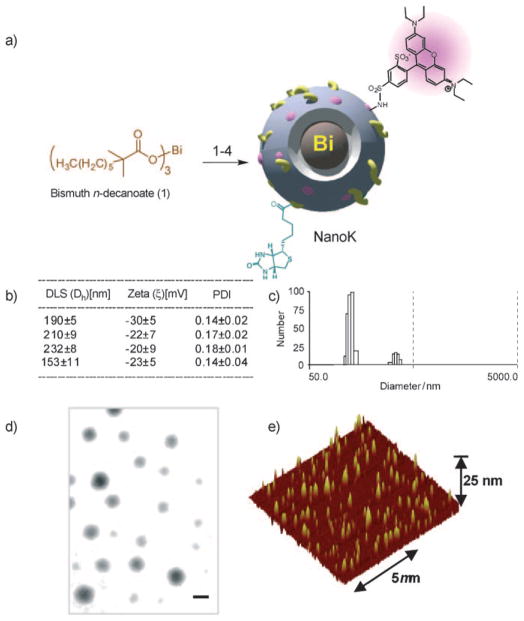
Pan Dipanjan, Roessl Ewald, Schlomka Jens-Peter, Caruthers Shelton D, Senpan Angana, Scott Mike J, Allen John S, Zhang Huiying, Hu Grace, Gaffney Patrick J, Choi Eric T, Rasche Volker, Wickline Samuel A, Proksa Roland, Lanza Gregory M

机构信息

C-TRAIN and Division of Cardiology, Washington University School of Medicine, 4320 Forest Park Avenue, Saint Louis, MO 63108, USA.

出版信息

Angew Chem Int Ed Engl. 2010 Dec 10;49(50):9635-9. doi: 10.1002/anie.201005657.

DOI:10.1002/anie.201005657
PMID:21077082
原文链接:https://pmc.ncbi.nlm.nih.gov/articles/PMC3096064/
Abstract
摘要
https://cdn.ncbi.nlm.nih.gov/pmc/blobs/34df/3096064/2447db718f4a/nihms282657f4.jpg
https://cdn.ncbi.nlm.nih.gov/pmc/blobs/34df/3096064/2e8741994859/nihms282657f1.jpg
https://cdn.ncbi.nlm.nih.gov/pmc/blobs/34df/3096064/591384cd070d/nihms282657f2.jpg
https://cdn.ncbi.nlm.nih.gov/pmc/blobs/34df/3096064/4842cfbdd8ce/nihms282657f3.jpg
https://cdn.ncbi.nlm.nih.gov/pmc/blobs/34df/3096064/2447db718f4a/nihms282657f4.jpg