Energy and Life Sciences Division, Midwest Research Institute, 425 Volker Boulevard, Kansas City, Missouri 64110, USA.
Virol J. 2011 Feb 11;8:66. doi: 10.1186/1743-422X-8-66.
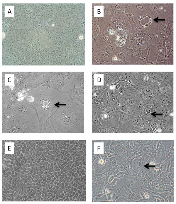
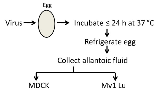
The infectivity of influenza A viruses can differ among the various primary cells and continuous cell lines used for such measurements. Over many years, we observed that all things equal, the cytopathic effects caused by influenza A subtype H1N1, H3N2, and H5N1 viruses were often detected earlier in a mink lung epithelial cell line (Mv1 Lu) than in MDCK cells. We asked whether virus yields as measured by the 50% tissue culture infectious dose and plaque forming titer also differed in MDCK and Mv1 Lu cells infected by the same influenza virus subtypes.
The 50% tissue culture infectious dose and plaque forming titer of many influenza A subtype H1N1, H3N2, and H5N1 viruses was higher in Mv1 Lu than in MDCK cells.
The yields of influenza subtype H1N1, H3N2, and H5N1 viruses can be higher in Mv1 Lu cells than in MDCK cells.
用于此类测量的各种原代细胞和连续细胞系中,甲型流感病毒的感染力可能存在差异。多年来,我们观察到,在所有条件相同的情况下,甲型 H1N1、H3N2 和 H5N1 亚型流感病毒引起的细胞病变效应通常在貂肺上皮细胞系(Mv1 Lu)中比在 MDCK 细胞中更早被检测到。我们想知道,当用相同的流感病毒亚型感染 MDCK 和 Mv1 Lu 细胞时,通过 50%组织培养感染剂量和噬菌斑形成滴度测量的病毒产量是否也不同。
许多甲型 H1N1、H3N2 和 H5N1 亚型流感病毒的 50%组织培养感染剂量和噬菌斑形成滴度在 Mv1 Lu 中的值均高于 MDCK 细胞。
甲型 H1N1、H3N2 和 H5N1 亚型流感病毒的产量在 Mv1 Lu 细胞中可能高于 MDCK 细胞。