Molecular Imaging Program, Center for Cancer Research, National Cancer Institute, NIH, Bethesda, MD 20892-1088, USA.
Contrast Media Mol Imaging. 2011 Jan-Feb;6(1):55-9. doi: 10.1002/cmmi.395. Epub 2010 May 28.
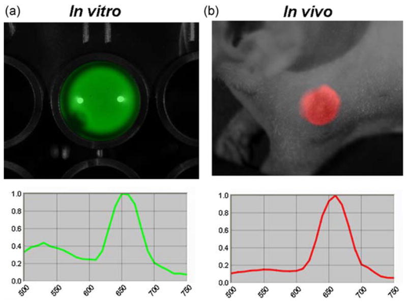
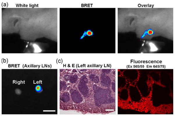
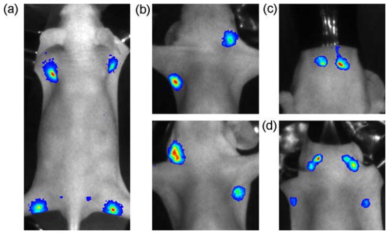
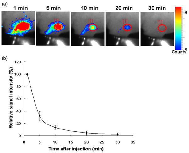
Autofluorescence arising from normal tissues can compromise the sensitivity and specificity of in vivo fluorescence imaging by lowering the target-to-background signal ratio. Since bioluminescence resonance energy transfer quantum dot (BRET-QDot) nano-particles can self-illuminate in near-infrared in the presence of the substrate, coelenterazine, without irradiating excitation lights, imaging using BRET-QDots does not produce any autofluorescence. In this study, we applied this BRET-QDot nano-particle to the in vivo lymphatic imaging in mice in order to compare with BRET, fluorescence or bioluminescence lymphatic imaging. BRET-QDot655, in which QDot655 is contained as a core, was injected at different sites (e.g. chin, ear, forepaws and hind paws) in mice followed by the intravenous coelenterazine injection, and then bioluminescence and fluorescence imaging were serially performed. In all mice, each lymphatic basin was clearly visualized in the BRET imaging with minimal background signals. The BRET signal in the lymph nodes lasted at least 30 min after coelenterazine injections. Furthermore, the BRET signal demonstrated better quantification than the fluorescence signal emitting from QDot655, the core of this BRET particle. These advantages of BRET-QDot allowed us to perform real-time, quantitative lymphatic imaging without image processing. BRET-Qdots have the potential to be a robust nano-material platform for developing optical molecular imaging probes.
自发荧光来自正常组织,可以通过降低靶标与背景信号的比率来降低体内荧光成像的灵敏度和特异性。由于生物发光共振能量转移量子点(BRET-QDot)纳米粒子在存在基质(腔肠素)的情况下可以在近红外光中自发光,而无需照射激发光,因此使用 BRET-QDot 进行成像不会产生任何自发荧光。在这项研究中,我们将这种 BRET-QDot 纳米粒子应用于小鼠体内淋巴成像,以便与 BRET、荧光或生物发光淋巴成像进行比较。BRET-QDot655 以 QDot655 为核心,在不同部位(如下巴、耳朵、前脚和后脚)注射小鼠体内,然后静脉注射腔肠素,然后连续进行生物发光和荧光成像。在所有小鼠中,BRET 成像中每个淋巴盆都清晰可见,背景信号最小。腔肠素注射后,淋巴结中的 BRET 信号至少持续 30 分钟。此外,BRET 信号比这种 BRET 粒子的核心 QDot655 发出的荧光信号具有更好的定量能力。BRET-QDot 的这些优势使我们能够进行实时、定量的淋巴成像,而无需图像处理。BRET-Qdots 有可能成为开发光学分子成像探针的强大纳米材料平台。