• 文献检索
  • 文档翻译
  • 深度研究
  • 学术资讯
  • Suppr Zotero 插件Zotero 插件
  • 邀请有礼
  • 套餐&价格
  • 历史记录
应用&插件
Suppr Zotero 插件Zotero 插件浏览器插件Mac 客户端Windows 客户端微信小程序
定价
高级版会员购买积分包购买API积分包
服务
文献检索文档翻译深度研究API 文档MCP 服务
关于我们
关于 Suppr公司介绍联系我们用户协议隐私条款
关注我们

Suppr 超能文献

核心技术专利:CN118964589B侵权必究
粤ICP备2023148730 号-1Suppr @ 2026

文献检索

告别复杂PubMed语法,用中文像聊天一样搜索,搜遍4000万医学文献。AI智能推荐,让科研检索更轻松。

立即免费搜索

文件翻译

保留排版,准确专业,支持PDF/Word/PPT等文件格式,支持 12+语言互译。

免费翻译文档

深度研究

AI帮你快速写综述,25分钟生成高质量综述,智能提取关键信息,辅助科研写作。

立即免费体验

体内微观光学投影断层成像。

Microscopic optical projection tomography in vivo.

机构信息

Institute of Molecular Biology and Biotechnology, Foundation for Research and Technology-Hellas, Crete, Greece.

出版信息

PLoS One. 2011 Apr 29;6(4):e18963. doi: 10.1371/journal.pone.0018963.

DOI:10.1371/journal.pone.0018963
PMID:21559481
原文链接:https://pmc.ncbi.nlm.nih.gov/articles/PMC3084718/
Abstract

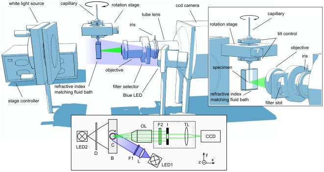
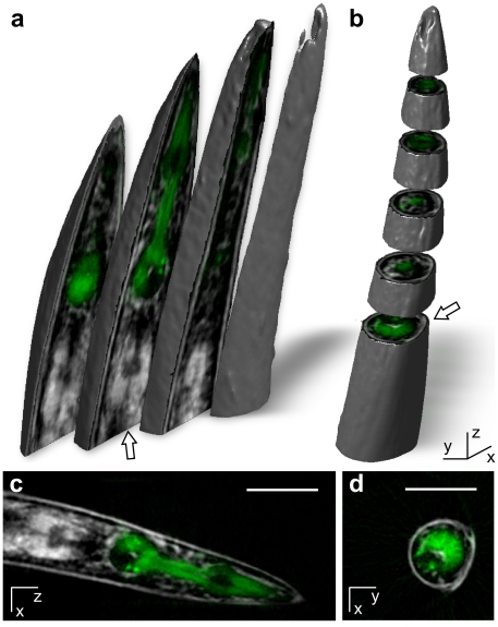
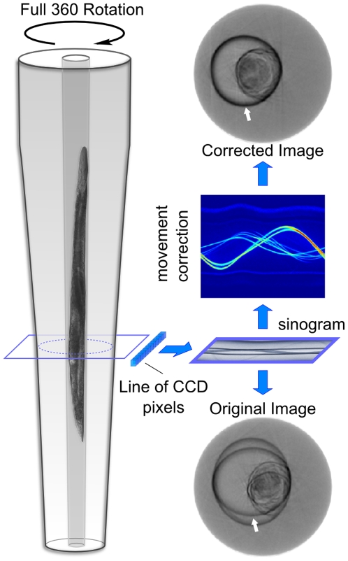
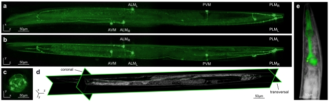
We describe a versatile optical projection tomography system for rapid three-dimensional imaging of microscopic specimens in vivo. Our tomographic setup eliminates the in xy and z strongly asymmetric resolution, resulting from optical sectioning in conventional confocal microscopy. It allows for robust, high resolution fluorescence as well as absorption imaging of live transparent invertebrate animals such as C. elegans. This system offers considerable advantages over currently available methods when imaging dynamic developmental processes and animal ageing; it permits monitoring of spatio-temporal gene expression and anatomical alterations with single-cell resolution, it utilizes both fluorescence and absorption as a source of contrast, and is easily adaptable for a range of small model organisms.

摘要

我们描述了一种多功能的光学投影层析成像系统,可用于快速对活体微观标本进行三维成像。我们的层析成像装置消除了传统共聚焦显微镜中光学切片在 xy 和 z 方向上产生的强烈不对称分辨率。它允许对活体透明无脊椎动物(如秀丽隐杆线虫)进行稳健的高分辨率荧光和吸收成像。与当前可用的方法相比,该系统在对动态发育过程和动物衰老进行成像时具有明显的优势;它可以以单细胞分辨率监测时空基因表达和解剖结构的改变,它同时利用荧光和吸收作为对比度的来源,并且易于适应一系列小型模式生物。

https://cdn.ncbi.nlm.nih.gov/pmc/blobs/915e/3084718/6fa571e3d0b0/pone.0018963.g004.jpg
https://cdn.ncbi.nlm.nih.gov/pmc/blobs/915e/3084718/2a345e9d9787/pone.0018963.g001.jpg
https://cdn.ncbi.nlm.nih.gov/pmc/blobs/915e/3084718/d4dd5861d010/pone.0018963.g002.jpg
https://cdn.ncbi.nlm.nih.gov/pmc/blobs/915e/3084718/57e7c560f3ca/pone.0018963.g003.jpg
https://cdn.ncbi.nlm.nih.gov/pmc/blobs/915e/3084718/6fa571e3d0b0/pone.0018963.g004.jpg

相似文献

1
Microscopic optical projection tomography in vivo.体内微观光学投影断层成像。
PLoS One. 2011 Apr 29;6(4):e18963. doi: 10.1371/journal.pone.0018963.
2
A customized light sheet microscope to measure spatio-temporal protein dynamics in small model organisms.一种用于测量小型模式生物中时空蛋白质动态的定制光片显微镜。
PLoS One. 2015 May 22;10(5):e0127869. doi: 10.1371/journal.pone.0127869. eCollection 2015.
3
Optical projection tomography.光学投影断层成像
Annu Rev Biomed Eng. 2004;6:209-28. doi: 10.1146/annurev.bioeng.6.040803.140210.
4
Tomographic imaging of macroscopic biomedical objects in high resolution and three dimensions using orthogonal-plane fluorescence optical sectioning.使用正交平面荧光光学切片技术对宏观生物医学物体进行高分辨率三维断层成像。
Appl Opt. 2009 Feb 10;48(5):941-8. doi: 10.1364/ao.48.000941.
5
Three-dimensional imaging of xenograft tumors using optical computed and emission tomography.利用光学计算机断层扫描和发射断层扫描对异种移植肿瘤进行三维成像。
Med Phys. 2006 Sep;33(9):3193-202. doi: 10.1118/1.2217109.
6
Noncontact optical imaging in mice with full angular coverage and automatic surface extraction.具有全角度覆盖和自动表面提取功能的小鼠非接触式光学成像。
Appl Opt. 2007 Jun 10;46(17):3617-27. doi: 10.1364/ao.46.003617.
7
Demonstrating Improved Multiple Transport-Mean-Free-Path Imaging Capabilities of Light Sheet Microscopy in the Quantification of Fluorescence Dynamics.演示光片显微镜在荧光动力学定量中的多重传输平均自由路径成像能力的改善。
Biotechnol J. 2018 Jan;13(1). doi: 10.1002/biot.201700419. Epub 2017 Dec 11.
8
Three-dimensional fluorescence-enhanced optical tomography using a hand-held probe based imaging system.使用基于手持探头成像系统的三维荧光增强光学层析成像技术。
Med Phys. 2008 Jul;35(7):3354-63. doi: 10.1118/1.2940603.
9
Free-space fluorescence molecular tomography utilizing 360 degrees geometry projections.利用360度几何投影的自由空间荧光分子断层成像。
Opt Lett. 2007 Feb 15;32(4):382-4. doi: 10.1364/ol.32.000382.
10
Transmission and fluorescence angular domain optical projection tomography of turbid media.浑浊介质的透射和荧光角域光学投影断层成像
Appl Opt. 2009 Nov 20;48(33):6448-57. doi: 10.1364/AO.48.006448.

引用本文的文献

1
Making sense of blobs, whorls, and shades: methods for label-free, inverse imaging in bright-field optical microscopy.理解斑点、漩涡和阴影:明场光学显微镜中无标记逆成像的方法。
Biophys Rev. 2025 Mar 18;17(2):335-345. doi: 10.1007/s12551-025-01301-1. eCollection 2025 Apr.
2
Reconstructing Three-Dimensional Optical Anisotropy with Tomographic Müller-Polarimetric Microscopy.用层析穆勒偏振显微镜重建三维光学各向异性
Adv Sci (Weinh). 2025 Jul;12(27):e2502075. doi: 10.1002/advs.202502075. Epub 2025 May 8.
3
Optical projection tomography implemented for accessibility and low cost ().

本文引用的文献

1
Correction for specimen movement and rotation errors for in-vivo Optical Projection Tomography.用于体内光学投影断层成像的样本移动和旋转误差校正
Biomed Opt Express. 2010 Jul 14;1(1):87-96. doi: 10.1364/BOE.1.000087.
2
Live optical projection tomography.实时光学投影断层成像。
Organogenesis. 2009 Oct;5(4):211-6. doi: 10.4161/org.5.4.10426.
3
Dual-modal three-dimensional imaging of single cells with isometric high resolution using an optical projection tomography microscope.使用光学投影断层显微镜对单细胞进行等距高分辨率的双模三维成像。
为实现可及性和低成本而实施的光学投影断层扫描()。 (括号部分原文缺失具体内容)
Philos Trans A Math Phys Eng Sci. 2024 Jul 9;382(2274):20230101. doi: 10.1098/rsta.2023.0101. Epub 2024 Jun 3.
4
Optical projection tomography reconstruction with few views using highly-generalizable deep learning at sinogram domain.在正弦图域使用高度通用的深度学习进行少视图光学投影断层扫描重建。
Biomed Opt Express. 2023 Nov 13;14(12):6260-6270. doi: 10.1364/BOE.500152. eCollection 2023 Dec 1.
5
Label-free optical imaging in developmental biology [Invited].发育生物学中的无标记光学成像[特邀文章]
Biomed Opt Express. 2020 Mar 13;11(4):2017-2040. doi: 10.1364/BOE.381359. eCollection 2020 Apr 1.
6
OptiJ: Open-source optical projection tomography of large organ samples.OptiJ:开源大型器官样本光学投影层析成像。
Sci Rep. 2019 Oct 30;9(1):15693. doi: 10.1038/s41598-019-52065-0.
7
Phase-Retrieved Tomography enables Mesoscopic imaging of Opaque Tumor Spheroids.相衬层析成像使不透明肿瘤球体的介观成像成为可能。
Sci Rep. 2017 Sep 19;7(1):11854. doi: 10.1038/s41598-017-12193-x.
8
Label-free three-dimensional imaging of Caenorhabditis elegans with visible optical coherence microscopy.利用可见光学相干显微镜对秀丽隐杆线虫进行无标记三维成像。
PLoS One. 2017 Jul 20;12(7):e0181676. doi: 10.1371/journal.pone.0181676. eCollection 2017.
9
Macro optical projection tomography for large scale 3D imaging of plant structures and gene activity.用于植物结构和基因活性大规模三维成像的宏观光学投影层析成像技术。
J Exp Bot. 2017 Jan 1;68(3):527-538. doi: 10.1093/jxb/erw452.
10
A hybrid microfluidic device for on-demand orientation and multidirectional imaging of organs and neurons.一种用于按需定向和对器官及神经元进行多方向成像的混合微流控装置。
Biomicrofluidics. 2016 Dec 1;10(6):064111. doi: 10.1063/1.4971157. eCollection 2016 Nov.
J Biomed Opt. 2009 Nov-Dec;14(6):064035. doi: 10.1117/1.3275470.
4
Microfluidics for the analysis of behavior, nerve regeneration, and neural cell biology in C. elegans.微流控技术在秀丽隐杆线虫行为分析、神经再生和神经细胞生物学中的应用。
Curr Opin Neurobiol. 2009 Oct;19(5):561-7. doi: 10.1016/j.conb.2009.10.010. Epub 2009 Nov 5.
5
Prohibitin couples diapause signalling to mitochondrial metabolism during ageing in C. elegans.在秀丽隐杆线虫衰老过程中,抑制素将滞育信号与线粒体代谢联系起来。
Nature. 2009 Oct 8;461(7265):793-7. doi: 10.1038/nature08466.
6
Fluorescence lifetime optical projection tomography.荧光寿命光学投影断层成像。
J Biophotonics. 2008 Oct;1(5):390-4. doi: 10.1002/jbio.200810044.
7
In vitro whole-organ imaging: 4D quantification of growing mouse limb buds.体外全器官成像:生长中小鼠肢芽的4D定量分析
Nat Methods. 2008 Jul;5(7):609-12. doi: 10.1038/nmeth.1219. Epub 2008 May 30.
8
Femtosecond laser nanoaxotomy lab-on-a-chip for in vivo nerve regeneration studies.用于体内神经再生研究的飞秒激光纳米轴突切断术芯片实验室
Nat Methods. 2008 Jun;5(6):531-3. doi: 10.1038/nmeth.1203. Epub 2008 Apr 13.
9
In vivo imaging of Drosophila melanogaster pupae with mesoscopic fluorescence tomography.利用介观荧光断层扫描对黑腹果蝇蛹进行体内成像。
Nat Methods. 2008 Jan;5(1):45-7. doi: 10.1038/nmeth1149. Epub 2007 Dec 9.
10
Microfluidics for in vivo imaging of neuronal and behavioral activity in Caenorhabditis elegans.用于秀丽隐杆线虫神经元和行为活动体内成像的微流控技术。
Nat Methods. 2007 Sep;4(9):727-31. doi: 10.1038/nmeth1075. Epub 2007 Aug 19.