Department of Chemistry and Chemical Biology, Harvard University, Cambridge, Massachusetts, United States of America.
PLoS One. 2011 May 6;6(5):e18940. doi: 10.1371/journal.pone.0018940.
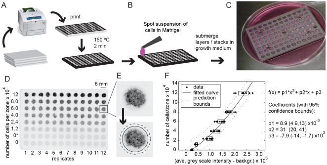
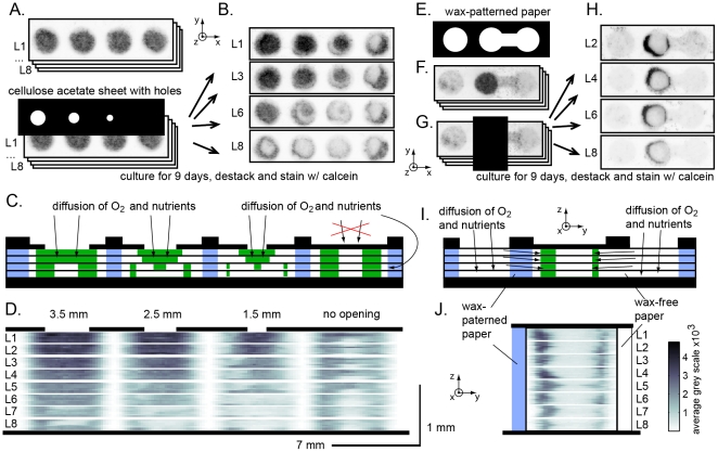
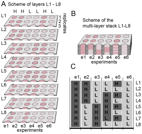
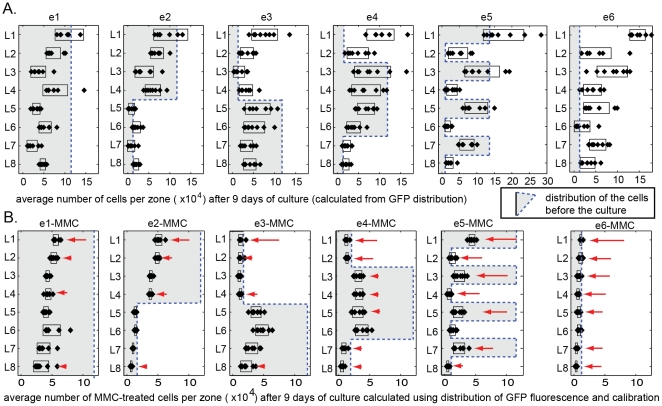
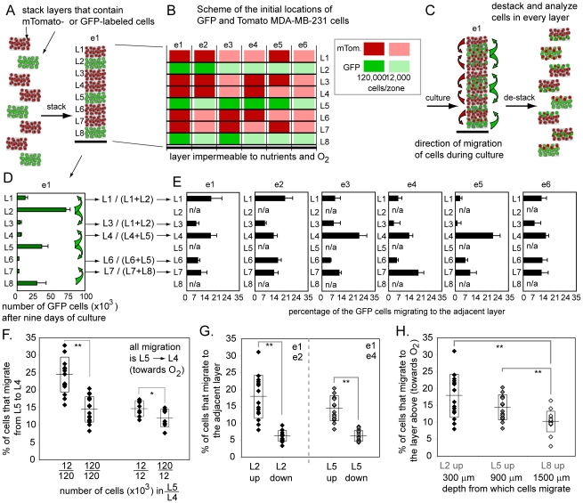
In vitro 3D culture is an important model for tissues in vivo. Cells in different locations of 3D tissues are physiologically different, because they are exposed to different concentrations of oxygen, nutrients, and signaling molecules, and to other environmental factors (temperature, mechanical stress, etc). The majority of high-throughput assays based on 3D cultures, however, can only detect the average behavior of cells in the whole 3D construct. Isolation of cells from specific regions of 3D cultures is possible, but relies on low-throughput techniques such as tissue sectioning and micromanipulation. Based on a procedure reported previously ("cells-in-gels-in-paper" or CiGiP), this paper describes a simple method for culture of arrays of thin planar sections of tissues, either alone or stacked to create more complex 3D tissue structures. This procedure starts with sheets of paper patterned with hydrophobic regions that form 96 hydrophilic zones. Serial spotting of cells suspended in extracellular matrix (ECM) gel onto the patterned paper creates an array of 200 micron-thick slabs of ECM gel (supported mechanically by cellulose fibers) containing cells. Stacking the sheets with zones aligned on top of one another assembles 96 3D multilayer constructs. De-stacking the layers of the 3D culture, by peeling apart the sheets of paper, "sections" all 96 cultures at once. It is, thus, simple to isolate 200-micron-thick cell-containing slabs from each 3D culture in the 96-zone array. Because the 3D cultures are assembled from multiple layers, the number of cells plated initially in each layer determines the spatial distribution of cells in the stacked 3D cultures. This capability made it possible to compare the growth of 3D tumor models of different spatial composition, and to examine the migration of cells in these structures.
体外 3D 培养是研究体内组织的重要模型。3D 组织中不同位置的细胞在生理上存在差异,因为它们暴露于不同浓度的氧气、营养物质和信号分子,以及其他环境因素(温度、机械应力等)。然而,大多数基于 3D 培养的高通量测定只能检测整个 3D 结构中细胞的平均行为。从 3D 培养物的特定区域分离细胞是可能的,但依赖于低通量技术,如组织切片和微操作。基于先前报道的一种程序(“细胞凝胶在纸上”或 CiGiP),本文描述了一种简单的方法,用于培养薄的组织平面切片阵列,无论是单独培养还是堆叠以创建更复杂的 3D 组织结构。该程序首先使用带有疏水区的纸张,疏水区形成 96 个亲水区域。将悬浮在细胞外基质 (ECM) 凝胶中的细胞连续点样到图案化的纸张上,形成含有细胞的 ECM 凝胶(由纤维素纤维机械支撑)的 200 微米厚的薄片阵列。通过将带有对齐区域的纸张堆叠在一起,组装 96 个 3D 多层结构。通过将 3D 培养物的层分层,将纸张剥离,“分离”96 个培养物中的所有“切片”。因此,可以简单地从 96 个区域阵列中的每个 3D 培养物中分离出 200 微米厚的含细胞薄片。由于 3D 培养物是由多个层组装而成的,因此每层最初接种的细胞数量决定了堆叠 3D 培养物中细胞的空间分布。这种能力使得比较不同空间组成的 3D 肿瘤模型的生长成为可能,并研究这些结构中细胞的迁移。