Department of Psychiatry and Psychotherapy, RWTH Aachen University, Aachen, Germany.
Neuroimage. 2011 Aug 1;57(3):938-49. doi: 10.1016/j.neuroimage.2011.05.021. Epub 2011 May 14.
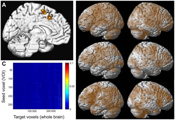
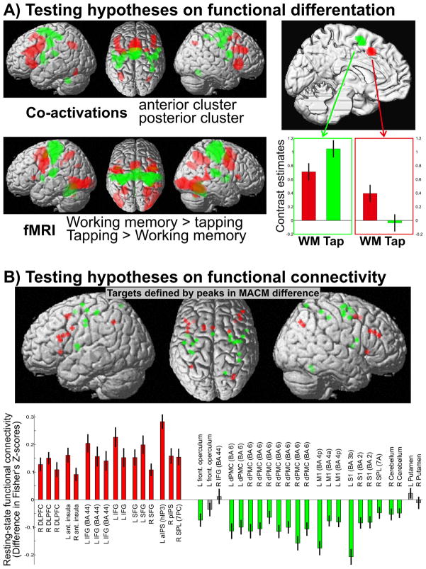
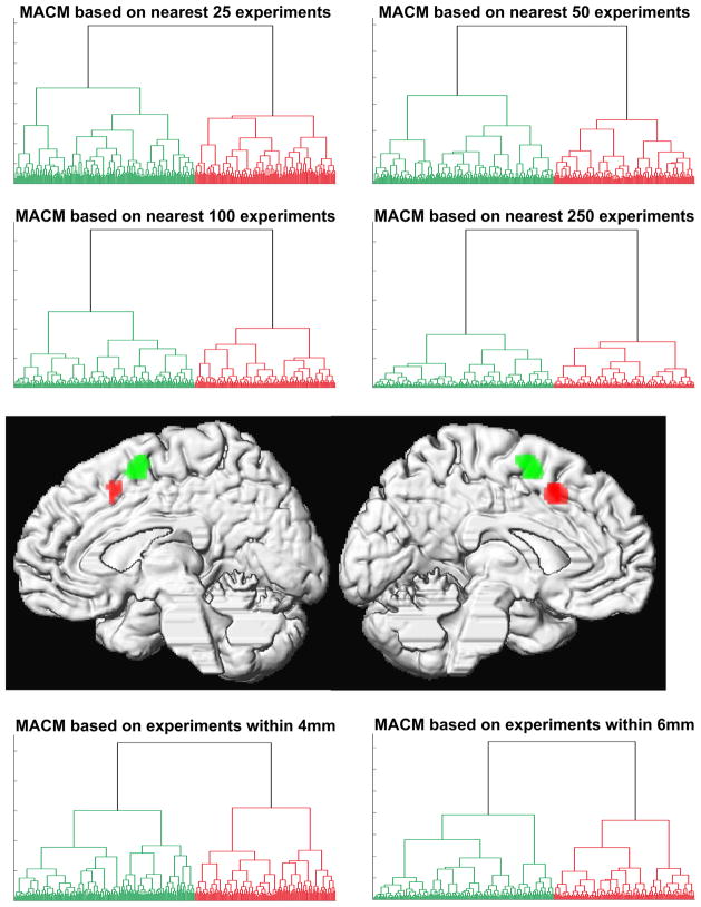
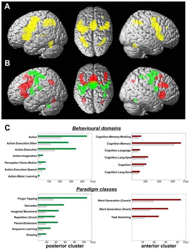
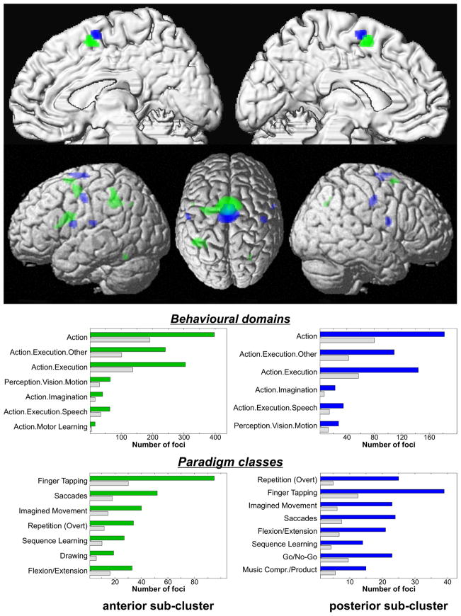
The organization of the cerebral cortex into distinct modules may be described along several dimensions, most importantly, structure, connectivity and function. Identification of cortical modules by differences in whole-brain connectivity profiles derived from diffusion tensor imaging or resting state correlations has already been shown. These approaches, however, carry no task-related information. Hence, inference on the functional relevance of the ensuing parcellation remains tentative. Here, we demonstrate, that Meta-Analytic Connectivity Modeling (MACM) allows the delineation of cortical modules based on their whole-brain co-activation pattern across databased neuroimaging results. Using a model free approach, two regions of the medial pre-motor cortex, SMA and pre-SMA were differentiated solely based on their functional connectivity. Assessing the behavioral domain and paradigm class meta-data of the experiments associated with the clusters derived from the co-activation based parcellation moreover allows the identification of their functional characteristics. The ensuing hypotheses about functional differentiation and distinct functional connectivity between pre-SMA and SMA were then explicitly tested and confirmed in independent datasets using functional and resting state fMRI. Co-activation based parcellation thus provides a new perspective for identifying modules of functional connectivity and linking them to functional properties, hereby generating new and subsequently testable hypotheses about the organization of cortical modules.
大脑皮层的组织可以沿着几个维度进行描述,最重要的是结构、连接和功能。已经证明,可以通过基于扩散张量成像或静息状态相关性的全脑连接谱的差异来识别皮质模块。然而,这些方法没有携带与任务相关的信息。因此,对随后分割的功能相关性的推断仍然是不确定的。在这里,我们证明,元分析连接建模(MACM)允许根据整个大脑在基于数据库的神经影像学结果中的共激活模式来描绘皮质模块。使用无模型方法,仅基于功能连接就可以区分内侧运动前皮质的两个区域,SMA 和前 SMA。评估与基于共激活的分割得出的簇相关的实验的行为域和范式类元数据,还可以识别它们的功能特征。随后,关于前 SMA 和 SMA 之间功能分化和不同功能连接的假设,然后在独立数据集上使用功能和静息状态 fMRI 进行了明确测试和验证。因此,基于共激活的分割为识别功能连接模块并将其与功能特性联系起来提供了新的视角,从而产生关于皮质模块组织的新的、随后可测试的假设。