Hutchings D L, van Drunen Littel-van den Hurk S, Babiuk L A
Agriculture Canada, Health of Animals Laboratory, Saskatoon, Saskatchewan.
J Virol. 1990 Oct;64(10):5114-22. doi: 10.1128/JVI.64.10.5114-5122.1990.
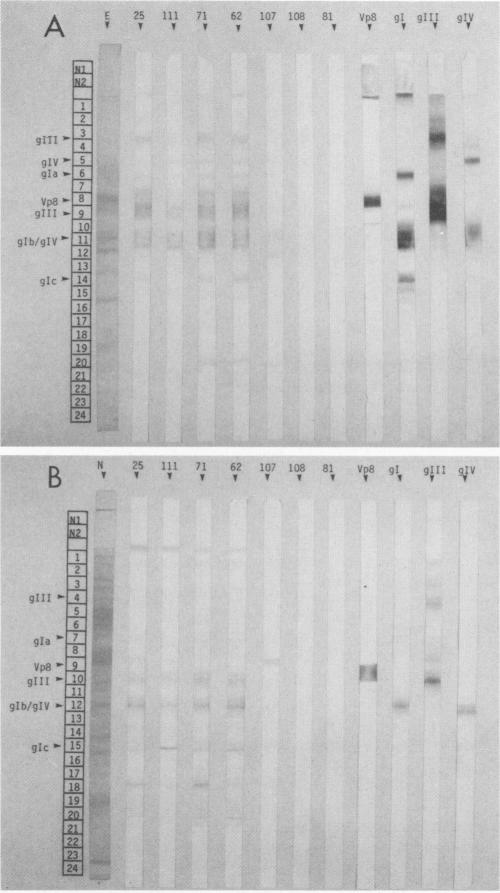
The immune response to bovine herpesvirus 1 (BHV-1) infection can protect cattle from subsequent challenge with the virus. This protection involves a variety of defensive strategies, and the activation of most of these defenses requires the recognition of viral proteins by the cellular immune system. To identify some of the BHV-1 proteins recognized by T lymphocytes, we measured in vitro proliferative responses to individual proteins. Viral proteins were separated by gel electrophoresis followed by Western immunoblotting, and immunoblots were evaluated for serological reactions. Unstained blotted fractions were processed into antigen-bearing particles for analysis in blastogenesis assays. Purified BHV-1 proteins obtained by immunoadsorbent chromatography were processed and included for comparison in both enzyme-linked immunosorbent and proliferation assays. The tegument protein VP8 and the glycoprotein gIV appeared to be the antigens which most consistently stimulated the proliferation of lymphocytes from BHV-1-immunized animals. Positive blastogenic responses were also detected to gI, gIII, and to one or more uncharacterized, low-molecular-weight proteins in some of the cattle tested. These results indicate that T-lymphocyte proliferative responses to BHV-1 proteins are detectable in immune cattle and may be important in protection from BHV-1 infection.
对牛疱疹病毒1型(BHV - 1)感染的免疫反应可保护牛免受该病毒随后的攻击。这种保护涉及多种防御策略,而这些防御机制中的大多数激活都需要细胞免疫系统识别病毒蛋白。为了鉴定一些被T淋巴细胞识别的BHV - 1蛋白,我们在体外测量了对单个蛋白的增殖反应。通过凝胶电泳分离病毒蛋白,随后进行Western免疫印迹,评估免疫印迹的血清学反应。将未染色的印迹部分加工成含抗原颗粒,用于在淋巴细胞增殖试验中进行分析。通过免疫吸附色谱法获得的纯化BHV - 1蛋白经过处理,并用于酶联免疫吸附试验和增殖试验中进行比较。被膜蛋白VP8和糖蛋白gIV似乎是最能持续刺激来自BHV - 1免疫动物淋巴细胞增殖的抗原。在一些测试的牛中,对gI、gIII以及一种或多种未鉴定的低分子量蛋白也检测到了阳性增殖反应。这些结果表明,在免疫牛中可检测到对BHV - 1蛋白的T淋巴细胞增殖反应,这可能在预防BHV - 1感染中起重要作用。