Department of Surgery, Stanford University, GK 201, Stanford, CA 94305, USA.
Biomaterials. 2012 Jan;33(1):80-90. doi: 10.1016/j.biomaterials.2011.09.041. Epub 2011 Oct 2.
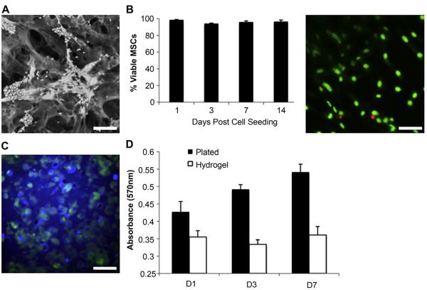
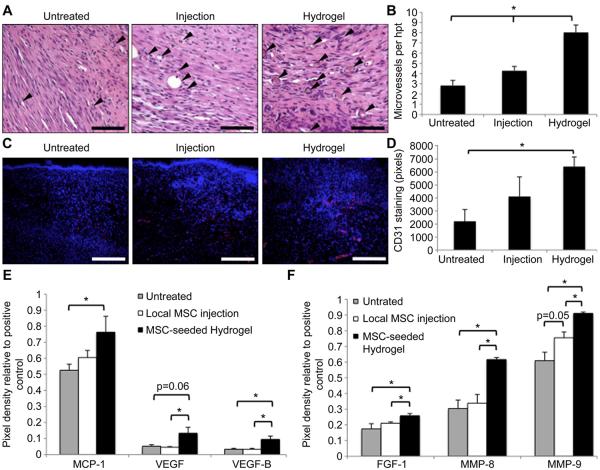
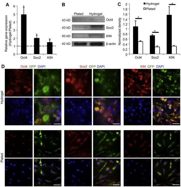
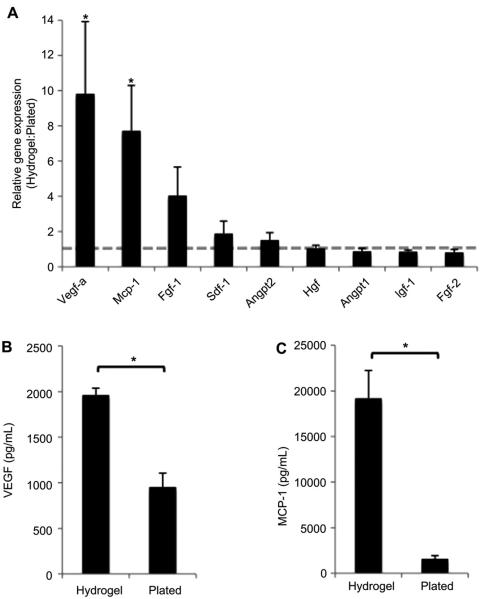
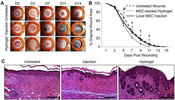
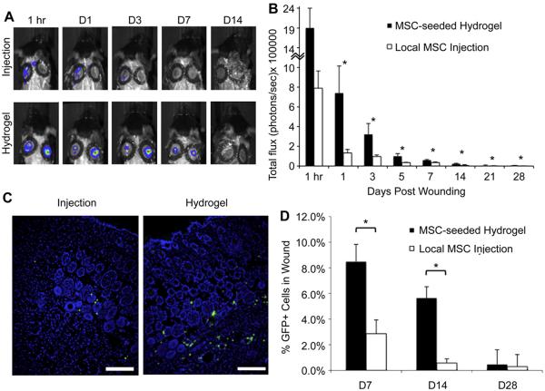
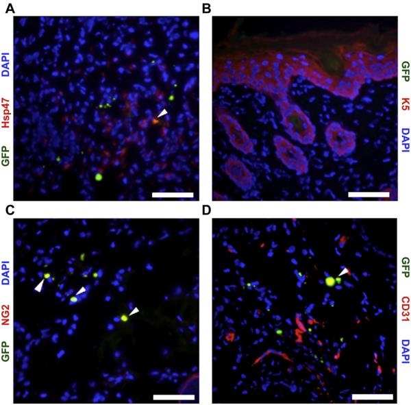
In this study, we examined the capacity of a biomimetic pullulan-collagen hydrogel to create a functional biomaterial-based stem cell niche for the delivery of mesenchymal stem cells (MSCs) into wounds. Murine bone marrow-derived MSCs were seeded into hydrogels and compared to MSCs grown in standard culture conditions. Hydrogels induced MSC secretion of angiogenic cytokines and expression of transcription factors associated with maintenance of pluripotency and self-renewal (Oct4, Sox2, Klf4) when compared to MSCs grown in standard conditions. An excisonal wound healing model was used to compare the ability of MSC-hydrogel constructs versus MSC injection alone to accelerate wound healing. Injection of MSCs did not significantly improve time to wound closure. In contrast, wounds treated with MSC-seeded hydrogels showed significantly accelerated healing and a return of skin appendages. Bioluminescence imaging and FACS analysis of luciferase+/GFP+ MSCs indicated that stem cells delivered within the hydrogel remained viable longer and demonstrated enhanced engraftment efficiency than those delivered via injection. Engrafted MSCs were found to differentiate into fibroblasts, pericytes and endothelial cells but did not contribute to the epidermis. Wounds treated with MSC-seeded hydrogels demonstrated significantly enhanced angiogenesis, which was associated with increased levels of VEGF and other angiogenic cytokines within the wounds. Our data suggest that biomimetic hydrogels provide a functional niche capable of augmenting MSC regenerative potential and enhancing wound healing.
在这项研究中,我们研究了仿生普鲁兰-胶原蛋白水凝胶在递送间充质干细胞(MSCs)进入伤口中创造功能性基于生物材料的干细胞龛的能力。将骨髓来源的鼠 MSCs 接种到水凝胶中,并与在标准培养条件下生长的 MSCs 进行比较。与在标准条件下生长的 MSCs 相比,水凝胶诱导 MSC 分泌血管生成细胞因子,并表达与维持多能性和自我更新相关的转录因子(Oct4、Sox2、Klf4)。使用切除性伤口愈合模型比较 MSC-水凝胶构建体与单独 MSC 注射加速伤口愈合的能力。MSC 注射并未显著缩短伤口愈合时间。相比之下,用 MSC 接种水凝胶处理的伤口显示出明显更快的愈合和皮肤附属物的恢复。荧光素酶+/GFP+ MSC 的生物发光成像和 FACS 分析表明,与通过注射递送的细胞相比,水凝胶内递送的干细胞具有更长的存活时间和增强的植入效率。植入的 MSCs 被发现分化为成纤维细胞、周细胞和内皮细胞,但不参与表皮形成。用 MSC 接种水凝胶处理的伤口显示出明显增强的血管生成,这与伤口内 VEGF 和其他血管生成细胞因子水平的增加有关。我们的数据表明,仿生水凝胶提供了一个功能性龛位,能够增强 MSC 的再生潜力并增强伤口愈合。