School of Biomedical Engineering Science and Health Systems, Drexel University, Philadelphia, PA, USA.
Acad Radiol. 2011 Nov;18(11):1341-8. doi: 10.1016/j.acra.2011.06.013.
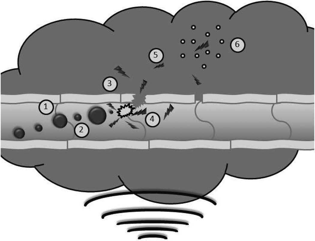
A doxorubicin-loaded microbubble has been developed that can be destroyed with focused ultrasound resulting in fragments, or "nanoshards" capable of escaping through the leaky tumor vasculature, promoting accumulation within the interstitium. This study uses a rat liver cancer model to examine the biodistribution and tumoral delivery of this microbubble platform compared with de novo drug-loaded polymer nanoparticles and free doxorubicin.
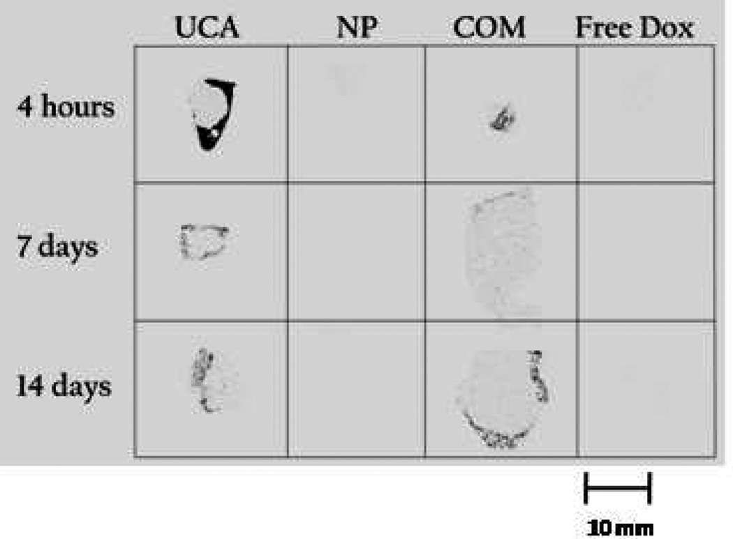
Microbubbles (1.8 μm) and 217-nm nanoparticles were prepared containing 14-C labeled doxorubicin. Microbubbles, nanoparticles, a combination of the two, or free doxorubicin were administered intravenously in rats bearing hepatomas, concomitant with tumor insonation. Doxorubicin levels in plasma, organs, and tumors were quantified after 4 hours and 7 and 14 days. Tumors were measured on sacrifice and evaluated with autoradiography and histology.
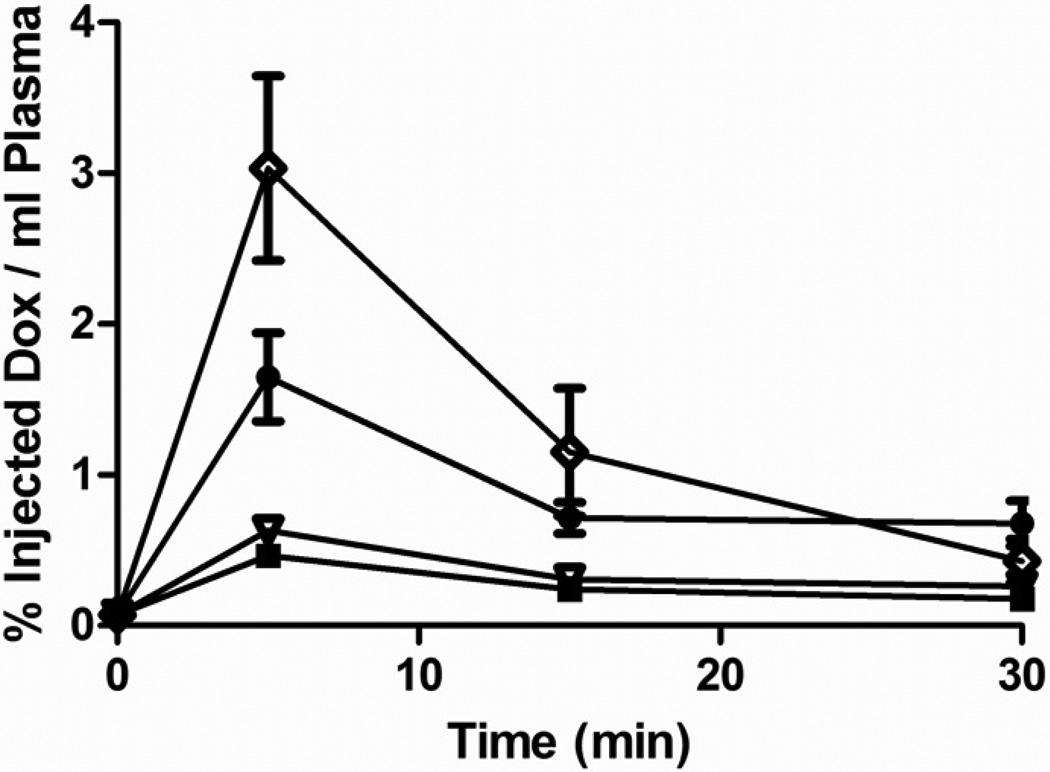
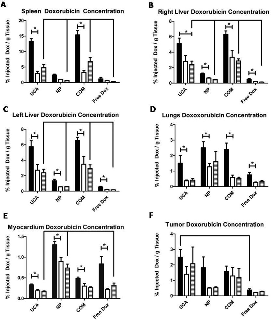
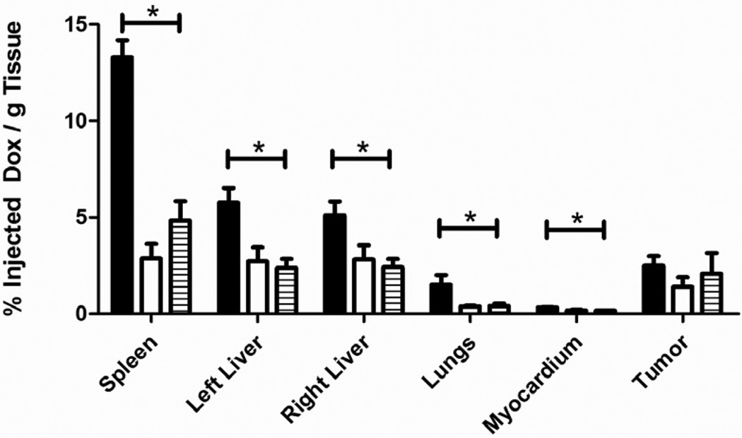
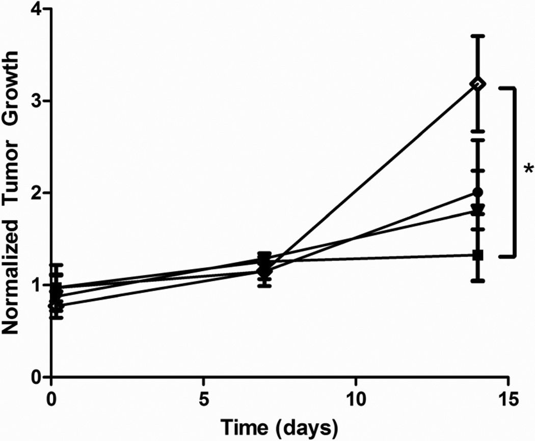
Animals treated with microbubbles had significantly lower plasma doxorubicin concentrations (0.466 ± 0.068%/mL) compared with free doxorubicin (3.033 ± 0.612%/mL, P = .0019). Drug levels in the myocardium were significantly lower in animals treated with microbubbles compared to free doxorubicin (0.168%/g tissue vs. 0.320%/g, P = .0088). Tumors treated with microbubbles showed significantly higher drug levels than tumors treated with free doxorubicin (2.491 ± 0.501 %/g vs. 0.373 ± 0.087 %/g, P = .0472). These tumors showed significantly less growth than tumors treated with free doxorubicin (P = .0390).
Doxorubicin loaded microbubbles triggered with ultrasound provided enhanced, sustained drug delivery to tumors, reduced plasma and myocardium doxorubicin levels, and arresting tumor growth. The results suggest that in situ generation of nano particles provides a superior treatment over injection of free drug and also de novo synthesized nanoparticles.
已经开发出一种载有阿霉素的微泡,通过聚焦超声破坏微泡会产生碎片,即“纳米碎片”,能够通过渗漏的肿瘤血管逃逸,促进在间质中的积累。本研究使用大鼠肝癌模型,比较了这种微泡平台与新合成的载药聚合物纳米颗粒和游离阿霉素的生物分布和肿瘤递送。
制备了含有 14-C 标记阿霉素的 1.8μm 微泡和 217nm 纳米颗粒。在荷肝癌大鼠中静脉注射微泡、纳米颗粒、两者的混合物或游离阿霉素,同时进行肿瘤超声照射。在 4 小时、7 天和 14 天后测量血浆、器官和肿瘤中的阿霉素水平。处死动物后测量肿瘤,并进行放射自显影和组织学评估。
与游离阿霉素(3.033 ± 0.612%/mL,P =.0019)相比,接受微泡治疗的动物血浆阿霉素浓度显著降低(0.466 ± 0.068%/mL)。与游离阿霉素(0.168%/g 组织 vs. 0.320%/g,P =.0088)相比,接受微泡治疗的动物心肌中的药物水平显著降低。接受微泡治疗的肿瘤与接受游离阿霉素治疗的肿瘤相比,药物水平显著升高(2.491 ± 0.501%/g vs. 0.373 ± 0.087%/g,P =.0472)。与接受游离阿霉素治疗的肿瘤相比,这些肿瘤的生长明显受到抑制(P =.0390)。
超声触发的载阿霉素微泡提供了增强的、持续的肿瘤药物输送,降低了血浆和心肌中的阿霉素水平,并抑制了肿瘤生长。结果表明,原位生成纳米颗粒比注射游离药物和新合成的纳米颗粒提供了更好的治疗效果。