Department of Chemistry and Volen Center for Complex Systems, Brandeis University, Waltham, MA, USA.
Expert Rev Proteomics. 2011 Oct;8(5):591-604. doi: 10.1586/epr.11.53.
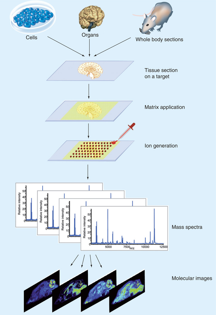
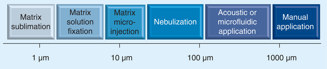
Single-cell analysis is gaining popularity in the field of mass spectrometry as a method for analyzing protein and peptide content in cells. The spatial resolution of MALDI mass spectrometry (MS) imaging is by a large extent limited by the laser focal diameter and the displacement of analytes during matrix deposition. Owing to recent advancements in both laser optics and matrix deposition methods, spatial resolution on the order of a single eukaryotic cell is now achievable by MALDI MS imaging. Provided adequate instrument sensitivity, a lateral resolution of approximately 10 µm is currently attainable with commercial instruments. As a result of these advances, MALDI MS imaging is poised to become a transformative clinical technology. In this article, the crucial steps needed to obtain single-cell resolution are discussed, as well as potential applications to disease research.
单细胞分析作为一种分析细胞内蛋白质和肽含量的方法,在质谱领域越来越受欢迎。MALDI 质谱 (MS) 成像的空间分辨率在很大程度上受到激光焦点直径和基质沉积过程中分析物位移的限制。由于激光光学和基质沉积方法的最新进展,现在通过 MALDI MS 成像可以实现单个真核细胞级别的空间分辨率。在仪器灵敏度足够的情况下,目前商用仪器可以达到大约 10 µm 的横向分辨率。由于这些进展,MALDI MS 成像有望成为一种变革性的临床技术。本文讨论了获得单细胞分辨率所需的关键步骤,以及其在疾病研究中的潜在应用。