Fondazione Matarelli, Dipartimento di Farmacologia, Chemioterapia e Tossicologia Medica, Università degli Studi di Milano, Milan, Italy.
Int J Nanomedicine. 2012;7:435-47. doi: 10.2147/IJN.S27537. Epub 2012 Jan 31.
We have previously shown that human mesenchymal stem cells (hMSCs) can reduce toxin-induced neurodegeneration in a well characterized rodent model of Parkinson's disease. However, the precise mechanisms, optimal cell concentration required for neuroprotection, and detailed cell tracking need to be defined. We exploited a near-infrared imaging platform to perform noninvasive tracing following transplantation of tagged hMSCs in live parkinsonian rats.
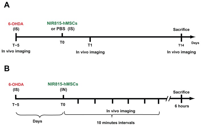
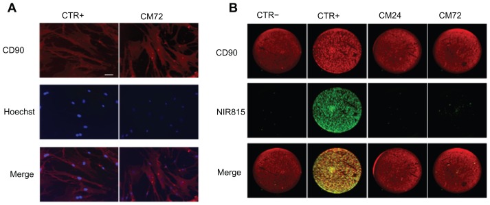
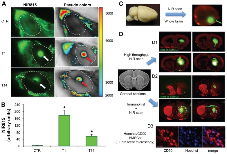
hMSCs were labeled both with a membrane intercalating dye, emitting in the near- infrared 815 nm spectrum, and the nuclear counterstain, Hoechst 33258. Effects of near-infrared dye on cell metabolism and proliferation were extensively evaluated in vitro. Tagged hMSCs were then administered to parkinsonian rats bearing a 6-hydroxydopamine-induced lesion of the nigrostriatal pathway, via two alternative routes, ie, intrastriatal or intranasal, and the cells were tracked in vivo and ex vivo using near-infrared technology.
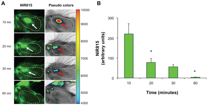
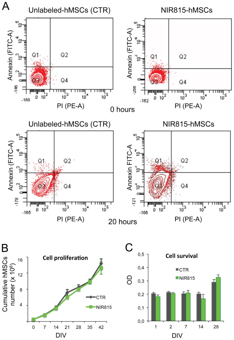
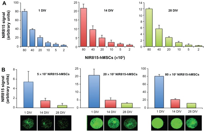
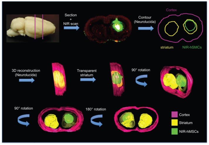
In vitro, NIR815 staining was stable in long-term hMSC cultures and did not interfere with cell metabolism or proliferation. A significant near-infrared signal was detectable in vivo, confined around the injection site for up to 14 days after intrastriatal transplantation. Conversely, following intranasal delivery, a strong near-infrared signal was immediately visible, but rapidly faded and was completely lost within 1 hour. After sacrifice, imaging data were confirmed by presence/absence of the Hoechst signal ex vivo in coronal brain sections. Semiquantitative analysis and precise localization of transplanted hMSCs were further performed ex vivo using near-infrared imaging.
Near-infrared technology allowed longitudinal detection of fluorescent-tagged cells in living animals giving immediate information on how different delivery routes affect cell distribution in the brain. Near-infrared imaging represents a valuable tool to evaluate multiple outcomes of transplanted cells, including their survival, localization, and migration over time within the host brain. This procedure considerably reduces the number of animal experiments needed, as well as interindividual variability, and may favor the development of efficient therapeutic strategies promptly applicable to patients.
我们之前已经证明,人骨髓间充质干细胞(hMSCs)可以减少帕金森病的一种特征明确的啮齿动物模型中毒素诱导的神经退行性变。然而,确切的机制、所需的最佳细胞浓度用于神经保护以及详细的细胞跟踪仍需确定。我们利用近红外成像平台在帕金森病大鼠活体中进行移植后标记 hMSC 的非侵入性跟踪。
hMSCs 同时用膜插入染料标记,发射近红外 815nm 光谱,并用核染料 Hoechst 33258 标记。在体外广泛评估近红外染料对细胞代谢和增殖的影响。然后,通过两种替代途径,即纹状体内或鼻内,将标记的 hMSC 施用于具有 6-羟多巴胺诱导的黑质纹状体通路损伤的帕金森病大鼠,并使用近红外技术在体内和体外进行跟踪。
在体外,NIR815 染色在长期 hMSC 培养中稳定,不会干扰细胞代谢或增殖。在纹状体内移植后,可在体内检测到长达 14 天的明显近红外信号,局限于注射部位周围。相反,鼻内给药后,立即出现强烈的近红外信号,但迅速消退,1 小时内完全消失。处死动物后,通过冠状脑切片的 Hoechst 信号的存在/不存在来确认成像数据。还使用近红外成像对移植的 hMSC 进行了半定量分析和精确的体外定位。
近红外技术允许在活体动物中对荧光标记细胞进行纵向检测,立即提供有关不同给药途径如何影响大脑中细胞分布的信息。近红外成像代表了一种有价值的工具,可用于评估移植细胞的多种结果,包括其在宿主大脑中的存活、定位和随时间的迁移。该程序大大减少了所需的动物实验数量以及个体间的变异性,并可能有利于迅速开发出适用于患者的有效治疗策略。