Rodrigues Eduardo B, Johanson Margara, Penha Fernando M
Department of Ophthalmology, Vision Institute, UNIFESP, Sao Paulo, Brazil.
J Ophthalmol. 2012;2012:806989. doi: 10.1155/2012/806989. Epub 2012 Jan 24.
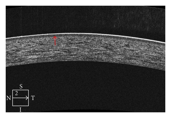
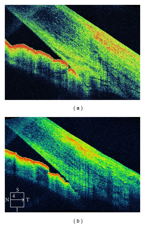
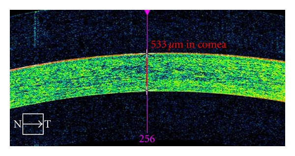
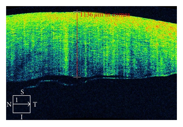
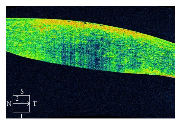
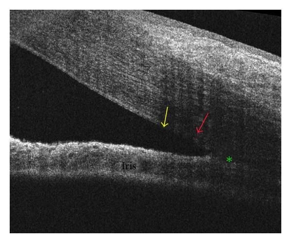
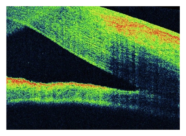
Optical coherence tomography (OCT) is an optical acquisition method to examine biological tissues. In recent years, OCT has become an important imaging technology used in diagnosing and following macular pathologies. Further development enabled application of optical coherence tomography in evaluation of the integrity of the nerve fiber layer, optic nerve cupping, anterior chamber angle, or corneal topography. In this manuscript we overview the use of OCT in the clinical practice to enable corneal, iris, ciliary body, and angle evaluation and diagnostics.
光学相干断层扫描(OCT)是一种用于检查生物组织的光学采集方法。近年来,OCT已成为诊断和跟踪黄斑病变的重要成像技术。进一步的发展使得光学相干断层扫描能够应用于评估神经纤维层的完整性、视神经杯、前房角或角膜地形图。在本手稿中,我们概述了OCT在临床实践中用于角膜、虹膜、睫状体和房角评估及诊断的应用。