• 文献检索
  • 文档翻译
  • 深度研究
  • 学术资讯
  • Suppr Zotero 插件Zotero 插件
  • 邀请有礼
  • 套餐&价格
  • 历史记录
应用&插件
Suppr Zotero 插件Zotero 插件浏览器插件Mac 客户端Windows 客户端微信小程序
定价
高级版会员购买积分包购买API积分包
服务
文献检索文档翻译深度研究API 文档MCP 服务
关于我们
关于 Suppr公司介绍联系我们用户协议隐私条款
关注我们

Suppr 超能文献

核心技术专利:CN118964589B侵权必究
粤ICP备2023148730 号-1Suppr @ 2026

文献检索

告别复杂PubMed语法,用中文像聊天一样搜索,搜遍4000万医学文献。AI智能推荐,让科研检索更轻松。

立即免费搜索

文件翻译

保留排版,准确专业,支持PDF/Word/PPT等文件格式,支持 12+语言互译。

免费翻译文档

深度研究

AI帮你快速写综述,25分钟生成高质量综述,智能提取关键信息,辅助科研写作。

立即免费体验

纳米多晶型羟基磷灰石涂层钛植入物的骨整合能力。

Bone integration capability of nanopolymorphic crystalline hydroxyapatite coated on titanium implants.

机构信息

Laboratory of Bone and Implant Sciences, The Weintraub Center for Reconstructive Biotechnology, Division of Advanced Prosthodontics, Biomaterials and Hospital Dentistry, UCLA School of Dentistry, Los Angeles, CA 90095-1668, USA.

出版信息

Int J Nanomedicine. 2012;7:859-73. doi: 10.2147/IJN.S28082. Epub 2012 Feb 17.

DOI:10.2147/IJN.S28082
PMID:22359461
原文链接:https://pmc.ncbi.nlm.nih.gov/articles/PMC3284227/
Abstract

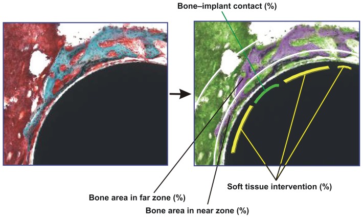
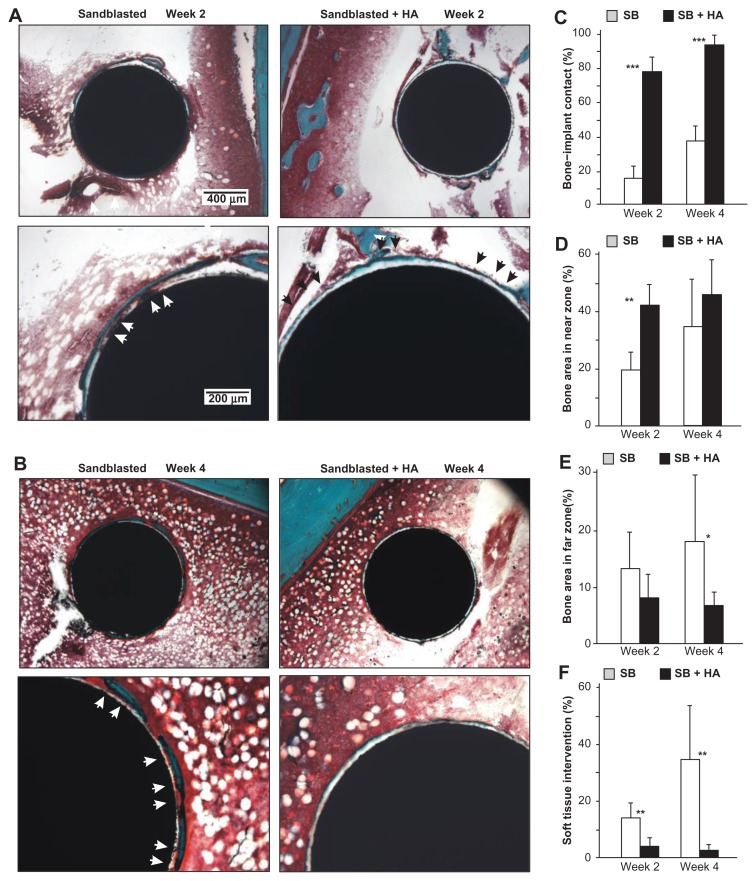
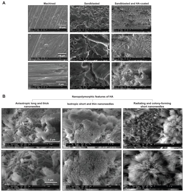
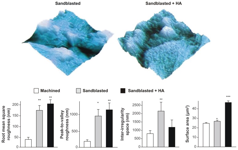
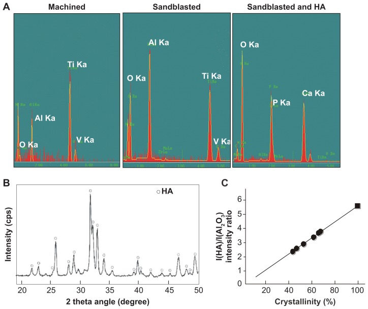
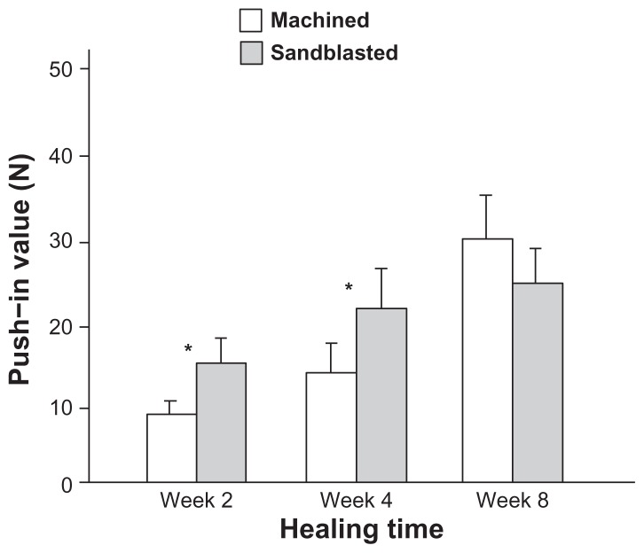
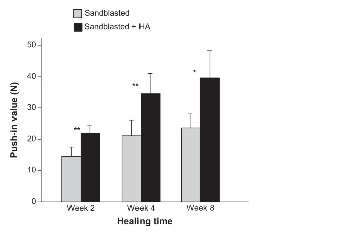
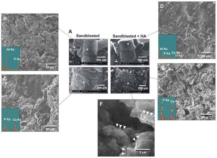
The mechanism by which hydroxyapatite (HA)-coated titanium promotes bone-implant integration is largely unknown. Furthermore, refining the fabrication of nano-structured HA to the level applicable to the mass production process for titanium implants is challenging. This study reports successful creation of nanopolymorphic crystalline HA on microroughened titanium surfaces using a combination of flame spray and low-temperature calcination and tests its biological capability to enhance bone-implant integration. Sandblasted microroughened titanium implants and sandblasted + HA-coated titanium implants were subjected to biomechanical and histomorphometric analyses in a rat model. The HA was 55% crystallized and consisted of nanoscale needle-like architectures developed in various diameters, lengths, and orientations, which resulted in a 70% increase in surface area compared to noncoated microroughened surfaces. The HA was free from impurity contaminants, with a calcium/phosphorus ratio of 1.66 being equivalent to that of stoichiometric HA. As compared to microroughened implants, HA-coated implants increased the strength of bone-implant integration consistently at both early and late stages of healing. HA-coated implants showed an increased percentage of bone-implant contact and bone volume within 50 μm proximity of the implant surface, as well as a remarkably reduced percentage of soft tissue intervention between bone and the implant surface. In contrast, bone volume outside the 50 μm border was lower around HA-coated implants. Thus, this study demonstrated that the addition of pure nanopolymorphic crystalline HA to microroughened titanium not only accelerates but also enhances the level of bone-implant integration and identified the specific tissue morphogenesis parameters modulated by HA coating. In particular, the nanocrystalline HA was proven to be drastic in increasing osteoconductivity and inhibiting soft tissue infiltration, but the effect was limited to the immediate microenvironment surrounding the implant.

摘要

羟基磷灰石(HA)涂层钛促进骨-植入物整合的机制在很大程度上尚不清楚。此外,将纳米结构 HA 的制造精化到适用于钛植入物大规模生产过程的水平具有挑战性。本研究报告了使用火焰喷涂和低温煅烧相结合的方法在微粗糙钛表面上成功制备纳米多晶型 HA,并测试了其增强骨-植入物整合的生物学能力。对喷砂微粗糙钛植入物和喷砂+HA 涂层钛植入物进行了生物力学和组织形态计量学分析,以大鼠模型进行了测试。HA 的结晶度为 55%,由纳米级针状结构组成,其直径、长度和方向各不相同,与非涂层微粗糙表面相比,表面积增加了 70%。HA 无杂质污染物,钙/磷比为 1.66,与化学计量的 HA 相当。与微粗糙植入物相比,HA 涂层植入物在愈合的早期和晚期均持续增加骨-植入物整合的强度。HA 涂层植入物增加了植入物表面附近 50 μm 范围内的骨-植入物接触和骨体积的百分比,同时显著降低了骨与植入物表面之间软组织介入的百分比。相比之下,HA 涂层植入物周围 50 μm 边界之外的骨体积较低。因此,本研究表明,将纯纳米多晶型 HA 添加到微粗糙钛中不仅可以加速,而且可以增强骨-植入物整合的水平,并确定了 HA 涂层调节的特定组织形态发生参数。特别是,纳米晶 HA 被证明在增加骨传导性和抑制软组织浸润方面非常有效,但效果仅限于植入物周围的直接微环境。

https://cdn.ncbi.nlm.nih.gov/pmc/blobs/f69d/3284227/c4f49057d75b/ijn-7-859f9.jpg
https://cdn.ncbi.nlm.nih.gov/pmc/blobs/f69d/3284227/2d159cad61b2/ijn-7-859f1.jpg
https://cdn.ncbi.nlm.nih.gov/pmc/blobs/f69d/3284227/add4b1c3765b/ijn-7-859f2.jpg
https://cdn.ncbi.nlm.nih.gov/pmc/blobs/f69d/3284227/3b20a1304afd/ijn-7-859f3.jpg
https://cdn.ncbi.nlm.nih.gov/pmc/blobs/f69d/3284227/09f15db388f1/ijn-7-859f4.jpg
https://cdn.ncbi.nlm.nih.gov/pmc/blobs/f69d/3284227/972725cbc76c/ijn-7-859f5.jpg
https://cdn.ncbi.nlm.nih.gov/pmc/blobs/f69d/3284227/6627c9bf8e13/ijn-7-859f6.jpg
https://cdn.ncbi.nlm.nih.gov/pmc/blobs/f69d/3284227/f4f202bc1709/ijn-7-859f7.jpg
https://cdn.ncbi.nlm.nih.gov/pmc/blobs/f69d/3284227/91f06da18a7e/ijn-7-859f8.jpg
https://cdn.ncbi.nlm.nih.gov/pmc/blobs/f69d/3284227/c4f49057d75b/ijn-7-859f9.jpg

相似文献

1
Bone integration capability of nanopolymorphic crystalline hydroxyapatite coated on titanium implants.纳米多晶型羟基磷灰石涂层钛植入物的骨整合能力。
Int J Nanomedicine. 2012;7:859-73. doi: 10.2147/IJN.S28082. Epub 2012 Feb 17.
2
Enhanced bone-integration capability of alkali- and heat-treated nanopolymorphic titanium in micro-to-nanoscale hierarchy.碱热处理纳米多晶钛在微纳分级结构中增强的骨整合能力。
Biomaterials. 2011 Oct;32(30):7297-308. doi: 10.1016/j.biomaterials.2011.06.033. Epub 2011 Jul 13.
3
Early-stage osseointegration capability of a submicrofeatured titanium surface created by microroughening and anodic oxidation.微粗糙化和阳极氧化处理构建的亚微米结构钛表面的早期骨整合能力。
Clin Oral Implants Res. 2013 Sep;24(9):991-1001. doi: 10.1111/j.1600-0501.2012.02507.x. Epub 2012 Jun 21.
4
A comparative study of zinc, magnesium, strontium-incorporated hydroxyapatite-coated titanium implants for osseointegration of osteopenic rats.锌、镁、锶掺杂羟基磷灰石涂层钛植入物促进骨质疏松大鼠骨整合的对比研究
Mater Sci Eng C Mater Biol Appl. 2016 May;62:226-32. doi: 10.1016/j.msec.2016.01.034. Epub 2016 Jan 16.
5
Bone integration capability of alkali- and heat-treated nanobimorphic Ti-15Mo-5Zr-3Al.碱热处理纳米晶 Ti-15Mo-5Zr-3Al 的骨整合能力。
Acta Biomater. 2011 Dec;7(12):4267-77. doi: 10.1016/j.actbio.2011.08.016. Epub 2011 Aug 24.
6
Bone integration capability of a series of strontium-containing hydroxyapatite coatings formed by micro-arc oxidation.微弧氧化形成的一系列含锶羟基磷灰石涂层的骨整合能力。
J Biomed Mater Res A. 2013 Sep;101(9):2465-80. doi: 10.1002/jbm.a.34548. Epub 2013 Jan 24.
7
The effect of magnesium-incorporated hydroxyapatite coating on titanium implant fixation in ovariectomized rats.镁掺杂羟基磷灰石涂层对去卵巢大鼠钛种植体固定的影响。
Int J Oral Maxillofac Implants. 2014 Jan-Feb;29(1):196-202. doi: 10.11607/jomi.2893.
8
The role of titanium implant surface modification with hydroxyapatite nanoparticles in progressive early bone-implant fixation in vivo.羟基磷灰石纳米颗粒改性钛种植体表面在体内渐进性早期骨-种植体固定中的作用。
Int J Oral Maxillofac Implants. 2009 Sep-Oct;24(5):808-16.
9
Biomechanical and morphometric analysis of hydroxyapatite-coated implants with varying crystallinity.不同结晶度的羟基磷灰石涂层植入物的生物力学和形态学分析。
J Oral Maxillofac Surg. 1999 Sep;57(9):1096-108; discussion 1108-9. doi: 10.1016/s0278-2391(99)90333-6.
10
Controlled Delivery of Curcumin and Vitamin K2 from Hydroxyapatite-Coated Titanium Implant for Enhanced in Vitro Chemoprevention, Osteogenesis, and in Vivo Osseointegration.载姜黄素和维生素 K2 的羟基磷灰石涂层钛植入物的控制释放用于增强体外化学预防、成骨作用和体内骨整合。
ACS Appl Mater Interfaces. 2020 Mar 25;12(12):13644-13656. doi: 10.1021/acsami.9b22474. Epub 2020 Mar 13.

引用本文的文献

1
The 3D theory of osseointegration: material, topography, and time as interdependent determinants of bone-implant integration.骨结合的三维理论:材料、表面形貌及时间作为骨与种植体结合的相互依存决定因素
Int J Implant Dent. 2025 Aug 2;11(1):49. doi: 10.1186/s40729-025-00639-1.
2
Nanotechnology in Orthopedic Care: Advances in Drug Delivery, Implants, and Biocompatibility Considerations.骨科护理中的纳米技术:药物递送、植入物及生物相容性考量方面的进展
Int J Nanomedicine. 2025 Jul 21;20:9251-9274. doi: 10.2147/IJN.S523462. eCollection 2025.
3
How Will Nanomedicine Revolutionize Future Dentistry and Periodontal Therapy?

本文引用的文献

1
Synergistic effects of UV photofunctionalization and micro-nano hybrid topography on the biological properties of titanium.UV 光功能化和微纳杂化形貌对钛生物学性能的协同效应。
Biomaterials. 2011 Jul;32(19):4358-68. doi: 10.1016/j.biomaterials.2011.03.001. Epub 2011 Mar 21.
2
On implant surfaces: a review of current knowledge and opinions.种植体表面:现有知识和观点的综述。
Int J Oral Maxillofac Implants. 2010 Jan-Feb;25(1):63-74.
3
Nanotopographical modification: a regulator of cellular function through focal adhesions.
纳米医学将如何彻底改变未来的牙科和牙周治疗?
Int J Mol Sci. 2025 Jan 12;26(2):592. doi: 10.3390/ijms26020592.
4
Nano-Based Approaches in Surface Modifications of Dental Implants: A Literature Review.基于纳米技术的牙种植体表面改性方法:文献综述。
Molecules. 2024 Jun 27;29(13):3061. doi: 10.3390/molecules29133061.
5
Beyond microroughness: novel approaches to navigate osteoblast activity on implant surfaces.超越微观粗糙度:探索在种植体表面调控成骨细胞活性的新方法。
Int J Implant Dent. 2024 Jul 5;10(1):35. doi: 10.1186/s40729-024-00554-x.
6
Genome-wide transcriptional responses of osteoblasts to different titanium surface topographies.成骨细胞对不同钛表面形貌的全基因组转录反应。
Mater Today Bio. 2023 Nov 4;23:100852. doi: 10.1016/j.mtbio.2023.100852. eCollection 2023 Dec.
7
The Effects of a Biomimetic Hybrid Meso- and Nano-Scale Surface Topography on Blood and Protein Recruitment in a Computational Fluid Dynamics Implant Model.仿生中尺度和纳米尺度表面形貌对计算流体动力学植入模型中血液和蛋白质募集的影响
Biomimetics (Basel). 2023 Aug 18;8(4):376. doi: 10.3390/biomimetics8040376.
8
Microenvironment-targeted strategy steers advanced bone regeneration.微环境靶向策略引领先进的骨再生。
Mater Today Bio. 2023 Jul 21;22:100741. doi: 10.1016/j.mtbio.2023.100741. eCollection 2023 Oct.
9
A biomechanical analysis of the effect of hydroxyapatite augmentation for trochanteric femoral fractures.羟基磷灰石增强术对股骨转子间骨折影响的生物力学分析
Exp Ther Med. 2023 Apr 18;25(6):256. doi: 10.3892/etm.2023.11955. eCollection 2023 Jun.
10
Ultraviolet Light Treatment of Titanium Microfiber Scaffolds Enhances Osteoblast Recruitment and Osteoconductivity in a Vertical Bone Augmentation Model: 3D UV Photofunctionalization.紫外线处理钛微纤维支架在垂直骨增量模型中增强成骨细胞募集和骨引导性:3D 紫外线光功能化。
Cells. 2022 Dec 21;12(1):19. doi: 10.3390/cells12010019.
纳米形貌修饰:通过黏着斑调节细胞功能。
Nanomedicine. 2010 Oct;6(5):619-33. doi: 10.1016/j.nano.2010.01.009. Epub 2010 Feb 4.
4
Selective cell affinity of biomimetic micro-nano-hybrid structured TiO2 overcomes the biological dilemma of osteoblasts.仿生微纳杂化结构 TiO2 的选择性细胞亲和性克服了成骨细胞的生物学困境。
Dent Mater. 2010 Apr;26(4):275-87. doi: 10.1016/j.dental.2009.11.077. Epub 2009 Dec 16.
5
Enhancement of bone-titanium integration profile with UV-photofunctionalized titanium in a gap healing model.在间隙愈合模型中,通过 UV 光功能化钛增强钛-骨整合特性。
Biomaterials. 2010 Mar;31(7):1546-57. doi: 10.1016/j.biomaterials.2009.11.018. Epub 2009 Dec 5.
6
Surface characterization of commercial oral implants on the nanometer level.商用口腔种植体的纳米级表面特性分析。
J Biomed Mater Res B Appl Biomater. 2010 Feb;92(2):462-9. doi: 10.1002/jbm.b.31538.
7
Effects of titanium surface topography on bone integration: a systematic review.钛表面形貌对骨整合的影响:一项系统评价
Clin Oral Implants Res. 2009 Sep;20 Suppl 4:172-84. doi: 10.1111/j.1600-0501.2009.01775.x.
8
Cellular behavior on TiO2 nanonodular structures in a micro-to-nanoscale hierarchy model.微米至纳米尺度层级模型中TiO₂纳米结节结构上的细胞行为
Biomaterials. 2009 Oct;30(29):5319-29. doi: 10.1016/j.biomaterials.2009.06.021. Epub 2009 Jul 9.
9
Nanotechnology for regenerative medicine.纳米技术在再生医学中的应用。
Biomed Microdevices. 2010 Aug;12(4):575-87. doi: 10.1007/s10544-008-9264-6.
10
The effect of ultraviolet functionalization of titanium on integration with bone.钛的紫外线功能化对与骨整合的影响。
Biomaterials. 2009 Feb;30(6):1015-25. doi: 10.1016/j.biomaterials.2008.11.004. Epub 2008 Nov 29.