• 文献检索
  • 文档翻译
  • 深度研究
  • 学术资讯
  • Suppr Zotero 插件Zotero 插件
  • 邀请有礼
  • 套餐&价格
  • 历史记录
应用&插件
Suppr Zotero 插件Zotero 插件浏览器插件Mac 客户端Windows 客户端微信小程序
定价
高级版会员购买积分包购买API积分包
服务
文献检索文档翻译深度研究API 文档MCP 服务
关于我们
关于 Suppr公司介绍联系我们用户协议隐私条款
关注我们

Suppr 超能文献

核心技术专利:CN118964589B侵权必究
粤ICP备2023148730 号-1Suppr @ 2026

文献检索

告别复杂PubMed语法,用中文像聊天一样搜索,搜遍4000万医学文献。AI智能推荐,让科研检索更轻松。

立即免费搜索

文件翻译

保留排版,准确专业,支持PDF/Word/PPT等文件格式,支持 12+语言互译。

免费翻译文档

深度研究

AI帮你快速写综述,25分钟生成高质量综述,智能提取关键信息,辅助科研写作。

立即免费体验

通过静电纺丝制备用于成骨细胞培养的仿生丝素蛋白/壳聚糖复合纳米纤维及其表征

Preparation and characterization of biomimetic silk fibroin/chitosan composite nanofibers by electrospinning for osteoblasts culture.

作者信息

Chen Jyh-Ping, Chen Shih-Hsien, Lai Guo-Jyun

机构信息

Department of Chemical and Materials Engineering, Chang Gung University, Kwei-Shan, Tao-Yuan, Taiwan, 333, Republic of China.

出版信息

Nanoscale Res Lett. 2012 Mar 6;7(1):170. doi: 10.1186/1556-276X-7-170.

DOI:10.1186/1556-276X-7-170
PMID:22394697
原文链接:https://pmc.ncbi.nlm.nih.gov/articles/PMC3310718/
Abstract

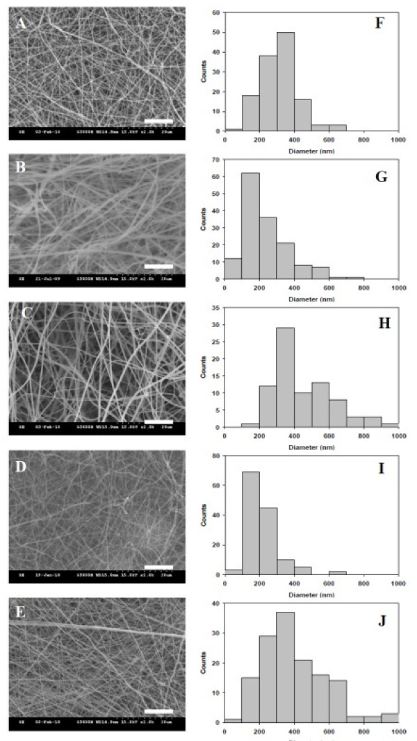
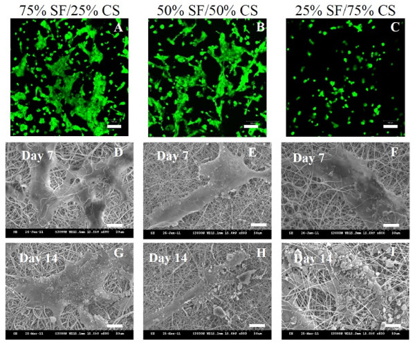
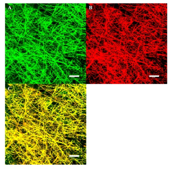
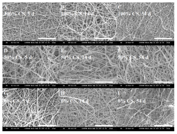
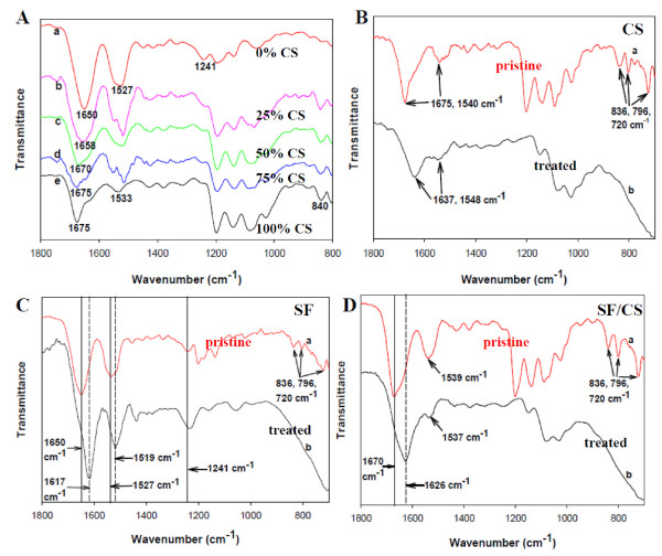
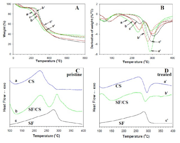
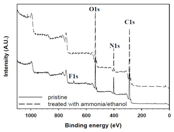
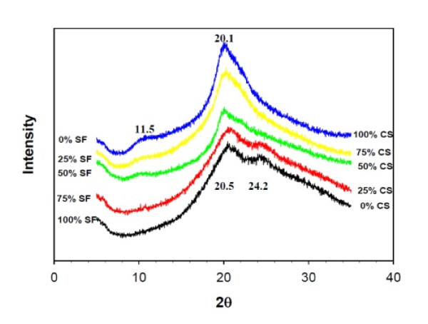
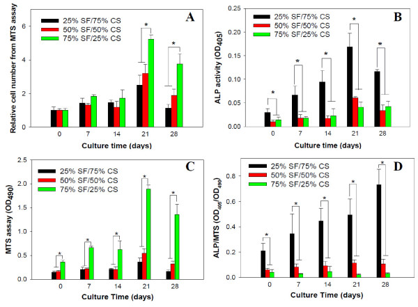
In this study, we have successfully fabricated electrospun bead-free silk fibroin [SF]/chitosan [CS] composite nanofibers [NFs] covering the whole range of CS content (0%, 25%, 50%, 75%, and 100%). SF/CS spinning solutions were prepared in a mixed solvent system of trifluoroacetic acid [TFA] and dichloromethane. The morphology of the NFs was observed by scanning electron microscope, and the average fiber diameter ranges from 215 to 478 nm. Confocal laser scanning microscopy confirms the uniform distribution of SF and CS within the composite NFs. To increase biocompatibility and preserve nanostructure when seeded with cells in culture medium, NFs were treated with an ethanol/ammonia aqueous solution to remove residual TFA and to change SF protein conformation. After the chemical treatment, SF/CS NFs could maintain the original structure for up to 54 days in culture medium. Properties of pristine and chemically treated SF/CS NFs were investigated by Fourier transform infrared spectroscopy [FT-IR], X-ray diffraction [XRD], and thermogravimetry/differential scanning calorimetry [TG/DSC]. Shift of absorption peaks in FT-IR spectra confirms the conformation change of SF from random coil to β-sheet by the action of ethanol, which is also consistent with the SF crystalline diffraction patterns measured by XRD. From TG/DSC analysis, the decomposition temperature peaks due to salt formation from TFA and protonated amines disappeared after chemical treatment, indicating complete removal of TFA by binding with ammonium ions during the treatment. This was also confirmed with the disappearance of F1s peak in X-ray photoelectron spectroscopy spectra and disappearance of TFA salt peaks in FT-IR spectra. The composite NFs could support the growth and osteogenic differentiation of human fetal osteoblastic [hFOB] cells, but each component in the composite NF shows distinct effect on cell behavior. SF promotes hFOB proliferation while CS enhances hFOB differentiation. The composite SF/CS NFs will be suitable for bone tissue engineering applications by choosing a suitable blend composition.PACS: 87.85.jf; 87.85.Rs; 68.37.Hk.

摘要

在本研究中,我们成功制备了覆盖壳聚糖(CS)全含量范围(0%、25%、50%、75%和100%)的无珠电纺丝素蛋白[SF]/壳聚糖[CS]复合纳米纤维[NFs]。SF/CS纺丝溶液在三氟乙酸[TFA]和二氯甲烷的混合溶剂体系中制备。通过扫描电子显微镜观察纳米纤维的形态,其平均纤维直径范围为215至478纳米。共聚焦激光扫描显微镜证实了SF和CS在复合纳米纤维中的均匀分布。为了在将细胞接种于培养基中时提高生物相容性并保留纳米结构,纳米纤维用乙醇/氨水溶液处理以去除残留的TFA并改变丝素蛋白的构象。化学处理后,SF/CS纳米纤维在培养基中最多可维持原始结构54天。通过傅里叶变换红外光谱[FT-IR]、X射线衍射[XRD]和热重分析/差示扫描量热法[TG/DSC]研究了原始和化学处理后的SF/CS纳米纤维的性能。FT-IR光谱中吸收峰的位移证实了乙醇作用下SF从无规卷曲构象转变为β-折叠构象,这也与XRD测量的丝素蛋白晶体衍射图谱一致。从TG/DSC分析可知,化学处理后由于TFA与质子化胺形成盐而产生的分解温度峰消失,表明处理过程中TFA通过与铵离子结合而被完全去除。这也通过X射线光电子能谱中F1s峰的消失以及FT-IR光谱中TFA盐峰的消失得到证实。复合纳米纤维能够支持人胎儿成骨细胞[hFOB]的生长和成骨分化,但复合纳米纤维中的每种成分对细胞行为都有不同的影响。SF促进hFOB增殖,而CS增强hFOB分化。通过选择合适的共混物组成,复合SF/CS纳米纤维将适用于骨组织工程应用。

PACS

87.85.jf;87.85.Rs;68.37.Hk。

https://cdn.ncbi.nlm.nih.gov/pmc/blobs/2b73/3310718/f8eac124eb97/1556-276X-7-170-9.jpg
https://cdn.ncbi.nlm.nih.gov/pmc/blobs/2b73/3310718/059d4e42c636/1556-276X-7-170-1.jpg
https://cdn.ncbi.nlm.nih.gov/pmc/blobs/2b73/3310718/5feb98cece77/1556-276X-7-170-2.jpg
https://cdn.ncbi.nlm.nih.gov/pmc/blobs/2b73/3310718/47f17976559b/1556-276X-7-170-3.jpg
https://cdn.ncbi.nlm.nih.gov/pmc/blobs/2b73/3310718/f4acee0d9df0/1556-276X-7-170-4.jpg
https://cdn.ncbi.nlm.nih.gov/pmc/blobs/2b73/3310718/248afaad88eb/1556-276X-7-170-5.jpg
https://cdn.ncbi.nlm.nih.gov/pmc/blobs/2b73/3310718/73d8a343ba48/1556-276X-7-170-6.jpg
https://cdn.ncbi.nlm.nih.gov/pmc/blobs/2b73/3310718/351dc0f71be8/1556-276X-7-170-7.jpg
https://cdn.ncbi.nlm.nih.gov/pmc/blobs/2b73/3310718/b3c3f33f15e3/1556-276X-7-170-8.jpg
https://cdn.ncbi.nlm.nih.gov/pmc/blobs/2b73/3310718/f8eac124eb97/1556-276X-7-170-9.jpg

相似文献

1
Preparation and characterization of biomimetic silk fibroin/chitosan composite nanofibers by electrospinning for osteoblasts culture.通过静电纺丝制备用于成骨细胞培养的仿生丝素蛋白/壳聚糖复合纳米纤维及其表征
Nanoscale Res Lett. 2012 Mar 6;7(1):170. doi: 10.1186/1556-276X-7-170.
2
Fabrication and intermolecular interactions of silk fibroin/hydroxybutyl chitosan blended nanofibers.丝素蛋白/羟丁基壳聚糖共混纳米纤维的制备及其分子间相互作用
Int J Mol Sci. 2011;12(4):2187-99. doi: 10.3390/ijms12042187. Epub 2011 Mar 30.
3
Production of silk sericin/silk fibroin blend nanofibers.丝胶/丝素共混纳米纤维的制备。
Nanoscale Res Lett. 2011 Aug 25;6(1):510. doi: 10.1186/1556-276X-6-510.
4
Response of human mesenchymal stem cells to intrafibrillar nanohydroxyapatite content and extrafibrillar nanohydroxyapatite in biomimetic chitosan/silk fibroin/nanohydroxyapatite nanofibrous membrane scaffolds.人骨髓间充质干细胞对仿生壳聚糖/丝素蛋白/纳米羟基磷灰石纳米纤维膜支架中纤维内纳米羟基磷灰石含量和纤维外纳米羟基磷灰石的反应
Int J Nanomedicine. 2015 Jan 12;10:567-84. doi: 10.2147/IJN.S73780. eCollection 2015.
5
Influence of Philosamia ricini silk fibroin components on morphology, secondary structure and thermal properties of chitosan biopolymer film.蓖麻蚕丝丝素蛋白成分对壳聚糖生物聚合物膜的形态、二级结构和热性能的影响
Pak J Biol Sci. 2009 Sep 15;12(18):1266-71. doi: 10.3923/pjbs.2009.1266.1271.
6
Composite chitosan/silk fibroin nanofibers for modulation of osteogenic differentiation and proliferation of human mesenchymal stem cells.用于调节人骨髓间充质干细胞成骨分化和增殖的复合壳聚糖/丝素纳米纤维。
Carbohydr Polym. 2014 Oct 13;111:288-97. doi: 10.1016/j.carbpol.2014.04.094. Epub 2014 May 4.
7
Electrospun poly (ɛ-caprolactone)/silk fibroin core-sheath nanofibers and their potential applications in tissue engineering and drug release.静电纺丝聚(己内酯)/丝素蛋白核壳纳米纤维及其在组织工程和药物释放中的潜在应用。
Int J Biol Macromol. 2011 Aug 1;49(2):223-32. doi: 10.1016/j.ijbiomac.2011.04.018. Epub 2011 May 5.
8
Preparation and characterization of genipin-cross-linked silk fibroin/chitosan sustained-release microspheres.京尼平交联丝素蛋白/壳聚糖缓释微球的制备与表征
Drug Des Devel Ther. 2015 May 4;9:2501-14. doi: 10.2147/DDDT.S78402. eCollection 2015.
9
Fabrication and Characterization of Silk Fibroin-Based Nanofibrous Scaffolds Supplemented with Gelatin for Corneal Tissue Engineering.基于丝素蛋白的纳米纤维支架的制备及特性研究——补充明胶用于角膜组织工程。
Cells Tissues Organs. 2021;210(3):173-194. doi: 10.1159/000515946. Epub 2021 Jul 12.
10
Characterization, Biocompatibility, and Optimization of Electrospun SF/PCL/CS Composite Nanofibers.静电纺丝SF/PCL/CS复合纳米纤维的表征、生物相容性及优化
Polymers (Basel). 2020 Jun 27;12(7):1439. doi: 10.3390/polym12071439.

引用本文的文献

1
Nano-scaffold-based delivery systems of antimicrobial agents in the treatment of osteomyelitis ; a narrative review.基于纳米支架的抗菌剂递送系统在骨髓炎治疗中的应用;一篇叙述性综述
Heliyon. 2024 Sep 24;10(21):e38392. doi: 10.1016/j.heliyon.2024.e38392. eCollection 2024 Nov 15.
2
Fabrication of Quercetin-Functionalized Morpholine and Pyridine Motifs-Laden Silk Fibroin Nanofibers for Effective Wound Healing in Preclinical Study.用于临床前研究中有效伤口愈合的槲皮素功能化含吗啉和吡啶基序的丝素蛋白纳米纤维的制备
Pharmaceutics. 2024 Mar 26;16(4):462. doi: 10.3390/pharmaceutics16040462.
3
Wet-Spun Chitosan-Sodium Caseinate Fibers for Biomedicine: From Spinning Process to Physical Properties.

本文引用的文献

1
Production of silk sericin/silk fibroin blend nanofibers.丝胶/丝素共混纳米纤维的制备。
Nanoscale Res Lett. 2011 Aug 25;6(1):510. doi: 10.1186/1556-276X-6-510.
2
Hydroxyapatite Mineralization on the Calcium Chloride Blended Polyurethane Nanofiber via Biomimetic Method.仿生法在氯化钙共混聚氨酯纳米纤维上的羟基磷灰石矿化。
Nanoscale Res Lett. 2010 Aug 19;6(1):2. doi: 10.1007/s11671-010-9737-4.
3
Surface modification of electrospun PLLA nanofibers by plasma treatment and cationized gelatin immobilization for cartilage tissue engineering.
用于生物医学的湿法纺丝壳聚糖-酪蛋白酸钠纤维:从纺丝工艺到物理性能。
Int J Mol Sci. 2024 Feb 1;25(3):1768. doi: 10.3390/ijms25031768.
4
An Overview of Scaffolds and Biomaterials for Skin Expansion and Soft Tissue Regeneration: Insights on Zinc and Magnesium as New Potential Key Elements.用于皮肤扩张和软组织再生的支架与生物材料概述:关于锌和镁作为新潜在关键元素的见解
Polymers (Basel). 2023 Sep 22;15(19):3854. doi: 10.3390/polym15193854.
5
Graphene Oxide-Coated Patterned Silk Fibroin Films Promote Cell Adhesion and Induce Cardiomyogenic Differentiation of Human Mesenchymal Stem Cells.氧化石墨烯涂层图案化丝素蛋白薄膜促进人骨髓间充质干细胞黏附和诱导其向心肌细胞分化。
Biomolecules. 2023 Jun 14;13(6):990. doi: 10.3390/biom13060990.
6
Study on the Incorporation of Chitosan Flakes in Electrospun Polycaprolactone Scaffolds.壳聚糖薄片掺入电纺聚己内酯支架的研究
Polymers (Basel). 2022 Apr 7;14(8):1496. doi: 10.3390/polym14081496.
7
Chitosan/Silk Fibroin Materials for Biomedical Applications-A Review.用于生物医学应用的壳聚糖/丝素蛋白材料——综述
Polymers (Basel). 2022 Mar 26;14(7):1343. doi: 10.3390/polym14071343.
8
Fibrous Polymer-Based Composites Obtained by Electrospinning for Bone Tissue Engineering.用于骨组织工程的静电纺丝法制备的纤维聚合物基复合材料
Polymers (Basel). 2021 Dec 28;14(1):96. doi: 10.3390/polym14010096.
9
Recombinant Human Bone Morphogenic Protein-2 Immobilized Fabrication of Magnesium Functionalized Injectable Hydrogels for Controlled-Delivery and Osteogenic Differentiation of Rat Bone Marrow-Derived Mesenchymal Stem Cells in Femoral Head Necrosis Repair.重组人骨形态发生蛋白-2固定化制备镁功能化可注射水凝胶用于股骨头坏死修复中大鼠骨髓间充质干细胞的控释与成骨分化
Front Cell Dev Biol. 2021 Nov 25;9:723789. doi: 10.3389/fcell.2021.723789. eCollection 2021.
10
Biomaterials for Soft Tissue Repair and Regeneration: A Focus on Italian Research in the Field.用于软组织修复与再生的生物材料:聚焦意大利在该领域的研究
Pharmaceutics. 2021 Aug 26;13(9):1341. doi: 10.3390/pharmaceutics13091341.
等离子体处理和阳离子化明胶固定化对电纺 PLLA 纳米纤维的表面改性及其在软骨组织工程中的应用。
Acta Biomater. 2011 Jan;7(1):234-43. doi: 10.1016/j.actbio.2010.08.015. Epub 2010 Aug 20.
4
Silk fibroin nanoparticles for cellular uptake and control release.丝素纳米粒子用于细胞摄取和控制释放。
Int J Pharm. 2010 Mar 30;388(1-2):242-50. doi: 10.1016/j.ijpharm.2009.12.052. Epub 2010 Jan 8.
5
Novel chitin and chitosan nanofibers in biomedical applications.用于生物医学应用的新型壳聚糖和壳聚糖纳米纤维。
Biotechnol Adv. 2010 Jan-Feb;28(1):142-50. doi: 10.1016/j.biotechadv.2009.11.001.
6
Electrospun silk biomaterial scaffolds for regenerative medicine.静电纺丝丝素生物材料支架用于再生医学。
Adv Drug Deliv Rev. 2009 Oct 5;61(12):988-1006. doi: 10.1016/j.addr.2009.07.005. Epub 2009 Jul 28.
7
Electrospun collagen-chitosan nanofiber: a biomimetic extracellular matrix for endothelial cell and smooth muscle cell.静电纺丝胶原-壳聚糖纳米纤维:一种模拟细胞外基质的内皮细胞和平滑肌细胞。
Acta Biomater. 2010 Feb;6(2):372-82. doi: 10.1016/j.actbio.2009.07.024. Epub 2009 Jul 25.
8
Silk as a Biomaterial.丝绸作为一种生物材料。
Prog Polym Sci. 2007;32(8-9):991-1007. doi: 10.1016/j.progpolymsci.2007.05.013.
9
Mesenchymal stem cell proliferation and differentiation on an injectable calcium phosphate-chitosan composite scaffold.间充质干细胞在可注射磷酸钙-壳聚糖复合支架上的增殖与分化
Biomaterials. 2009 May;30(14):2675-82. doi: 10.1016/j.biomaterials.2009.01.022. Epub 2009 Feb 1.
10
Biological basis of bone formation, remodeling, and repair-part II: extracellular matrix.骨形成、重塑和修复的生物学基础——第二部分:细胞外基质
Tissue Eng Part B Rev. 2008 Sep;14(3):275-83. doi: 10.1089/ten.teb.2008.0083.