Beretta Mario, Cicciù Marco, Bramanti Ennio, Maiorana Carlo
Implantology Department, School of Dentistry, University of Milan, IRCSS Cà Grande, MI, Italy.
Int J Dent. 2012;2012:261905. doi: 10.1155/2012/261905. Epub 2012 Jul 17.
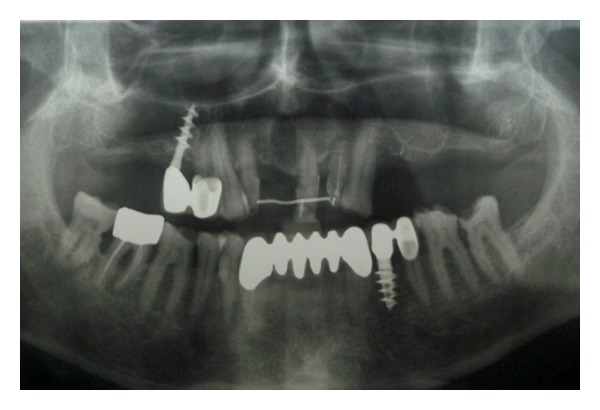
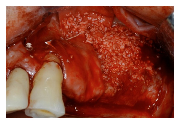
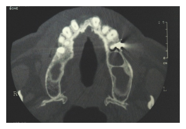
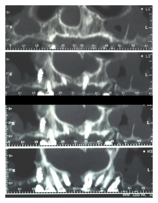
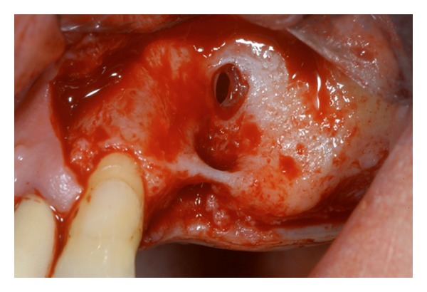
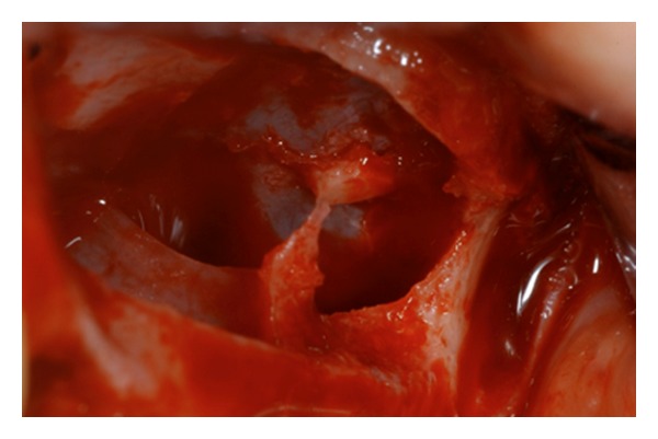
Maxillary sinus floor elevation via a lateral approach is a predictable technique to increase bone volume of the edentulous posterior maxilla and consequently for dental implants placement. The sinus floor is elevated and it can be augmented with either autologous or xenogeneic bone grafts following an opening bone window created on the facial buccal wall. Maxillary septa are walls of cortical bone within the maxillary sinus. The septa shape has been described as an inverted gothic arch arising from the inferior or lateral walls of the sinus and may even divide the sinus into two or more cavities. Some authors have reported a higher prevalence of septa in atrophic edentulous areas than in nonatrophic ones. Radiographic identification of these structures is important in order to perform the right design of the lateral window during sinus lift. Aim of this investigation is to highlight the correct steps for doing sinus lift surgery in presence of those anatomic variations. Clinicians should always perform clinical and radiographic diagnosis in order to avoid complications related to the sinus lift surgery.
经外侧入路的上颌窦底提升术是一种可预测的技术,用于增加无牙后上颌骨的骨量,从而便于植入牙种植体。上颌窦底被提升,在颊侧骨壁上创建一个开窗骨窗后,可以使用自体或异种骨移植材料进行增骨。上颌间隔是上颌窦内的皮质骨壁。间隔的形状被描述为从窦的下壁或侧壁产生的倒哥特式拱门,甚至可能将窦分成两个或更多个腔。一些作者报告说,萎缩性无牙区的间隔患病率高于非萎缩性无牙区。为了在窦提升术中正确设计外侧窗口,对这些结构进行影像学识别很重要。本研究的目的是强调在存在这些解剖变异的情况下进行窦提升手术的正确步骤。临床医生应始终进行临床和影像学诊断,以避免与窦提升手术相关的并发症。