• 文献检索
  • 文档翻译
  • 深度研究
  • 学术资讯
  • Suppr Zotero 插件Zotero 插件
  • 邀请有礼
  • 套餐&价格
  • 历史记录
应用&插件
Suppr Zotero 插件Zotero 插件浏览器插件Mac 客户端Windows 客户端微信小程序
定价
高级版会员购买积分包购买API积分包
服务
文献检索文档翻译深度研究API 文档MCP 服务
关于我们
关于 Suppr公司介绍联系我们用户协议隐私条款
关注我们

Suppr 超能文献

核心技术专利:CN118964589B侵权必究
粤ICP备2023148730 号-1Suppr @ 2026

文献检索

告别复杂PubMed语法,用中文像聊天一样搜索,搜遍4000万医学文献。AI智能推荐,让科研检索更轻松。

立即免费搜索

文件翻译

保留排版,准确专业,支持PDF/Word/PPT等文件格式,支持 12+语言互译。

免费翻译文档

深度研究

AI帮你快速写综述,25分钟生成高质量综述,智能提取关键信息,辅助科研写作。

立即免费体验

利用台式X射线相衬断层扫描和临界点干燥技术对组织微观结构进行无损体积成像。

Nondestructive volumetric imaging of tissue microstructure with benchtop x-ray phase-contrast tomography and critical point drying.

作者信息

Zysk Adam M, Garson Alfred B, Xu Qiaofeng, Brey Eric M, Zhou Wei, Brankov Jovan G, Wernick Miles N, Kuszak Jerome R, Anastasio Mark A

出版信息

Biomed Opt Express. 2012 Aug 1;3(8):1924-32. doi: 10.1364/BOE.3.001924. Epub 2012 Jul 24.

DOI:10.1364/BOE.3.001924
PMID:22876355
原文链接:https://pmc.ncbi.nlm.nih.gov/articles/PMC3409710/
Abstract

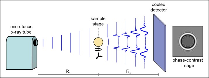
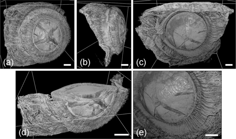
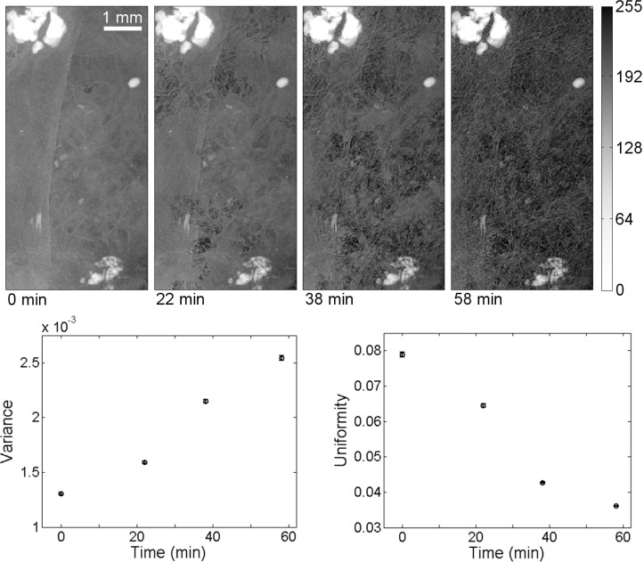
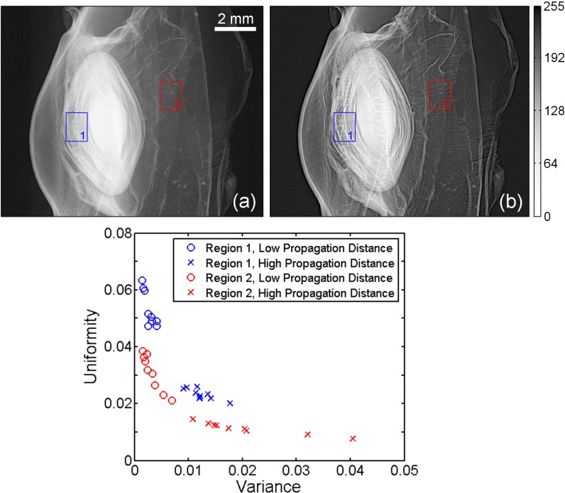
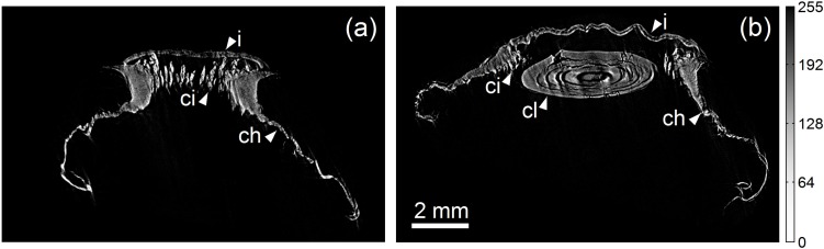
The in vitro investigation of many optically opaque biological microstructures requires 3D analysis at high resolution over a large field of view. We demonstrate a new nondestructive volumetric imaging technique that eliminates the structural and computational limitations of conventional 2D optical microscopy by combining x-ray phase-contrast tomography with critical point drying sample preparation. We experimentally demonstrate the enhancement of small features afforded by phase-contrast imaging and show the contrast improvement afforded by the drying of a hydrated specimen. We further demonstrate the biological application of this technique by imaging the microstructure of the accommodative apparatus in a primate eye using a benchtop phase-contrast tomography system.

摘要

对许多光学不透明的生物微观结构进行体外研究需要在大视野范围内进行高分辨率的三维分析。我们展示了一种新的无损体积成像技术,该技术通过将X射线相衬断层扫描与临界点干燥样品制备相结合,消除了传统二维光学显微镜的结构和计算限制。我们通过实验证明了相衬成像对小特征的增强作用,并展示了水合标本干燥所带来的对比度改善。我们还使用台式相衬断层扫描系统对灵长类动物眼睛中调节装置的微观结构进行成像,进一步证明了该技术的生物学应用。

https://cdn.ncbi.nlm.nih.gov/pmc/blobs/d172/3409710/b40c4c467247/boe-3-8-1924-g005.jpg
https://cdn.ncbi.nlm.nih.gov/pmc/blobs/d172/3409710/41f8445273c9/boe-3-8-1924-g001.jpg
https://cdn.ncbi.nlm.nih.gov/pmc/blobs/d172/3409710/efb830e09bc5/boe-3-8-1924-g002.jpg
https://cdn.ncbi.nlm.nih.gov/pmc/blobs/d172/3409710/7e071aa43ec1/boe-3-8-1924-g003.jpg
https://cdn.ncbi.nlm.nih.gov/pmc/blobs/d172/3409710/6cadde752702/boe-3-8-1924-g004.jpg
https://cdn.ncbi.nlm.nih.gov/pmc/blobs/d172/3409710/b40c4c467247/boe-3-8-1924-g005.jpg

相似文献

1
Nondestructive volumetric imaging of tissue microstructure with benchtop x-ray phase-contrast tomography and critical point drying.利用台式X射线相衬断层扫描和临界点干燥技术对组织微观结构进行无损体积成像。
Biomed Opt Express. 2012 Aug 1;3(8):1924-32. doi: 10.1364/BOE.3.001924. Epub 2012 Jul 24.
2
Three-dimensional imaging of whole mouse models: comparing nondestructive X-ray phase-contrast micro-CT with cryotome-based planar epi-illumination imaging.全鼠模型的三维成像:比较无损 X 射线相衬显微 CT 与基于冷冻切片的平面反射光照明成像。
J Microsc. 2014 Jan;253(1):24-30. doi: 10.1111/jmi.12094. Epub 2013 Nov 15.
3
A review of techniques for visualising soft tissue microstructure deformation and quantifying strain Ex Vivo.软组织微观结构变形可视化技术及体外应变定量评估研究综述。
J Microsc. 2018 Dec;272(3):165-179. doi: 10.1111/jmi.12701. Epub 2018 Apr 14.
4
Advantages of intermediate X-ray energies in Zernike phase contrast X-ray microscopy.中间 X 射线能量在泽尼克相衬 X 射线显微镜中的优势。
Biotechnol Adv. 2013 May-Jun;31(3):387-92. doi: 10.1016/j.biotechadv.2012.04.001. Epub 2012 Apr 13.
5
Three-Dimensional High-Resolution Digital Inline Hologram Reconstruction with a Volumetric Deconvolution Method.基于体三维反卷积的高分辨率数字全息三维重建。
Sensors (Basel). 2018 Sep 3;18(9):2918. doi: 10.3390/s18092918.
6
X-ray phase-contrast tomography of cells manipulated with an optical stretcher.用光学镊子操控的细胞的X射线相衬断层扫描术。
J Synchrotron Radiat. 2024 Jul 1;31(Pt 4):923-935. doi: 10.1107/S1600577524003618. Epub 2024 Jun 11.
7
Nanoscale Three-Dimensional Microstructural Characterization of an Sn-Rich Solder Alloy Using High-Resolution Transmission X-Ray Microscopy (TXM).使用高分辨率透射X射线显微镜(TXM)对富锡焊料合金进行纳米级三维微观结构表征。
Microsc Microanal. 2016 Aug;22(4):808-13. doi: 10.1017/S1431927616011429. Epub 2016 Jul 18.
8
Dark-Field Imaging: Recent developments and potential clinical applications.暗场成像:最新进展与潜在临床应用
Phys Med. 2016 Dec;32(12):1801-1812. doi: 10.1016/j.ejmp.2016.11.103.
9
Micron-scale 3D imaging of wood and plant microstructure using high-resolution X-ray phase-contrast microtomography.使用高分辨率 X 射线相衬显微断层摄影术对木材和植物微观结构进行微米级 3D 成像。
J Struct Biol. 2010 Aug;171(2):182-8. doi: 10.1016/j.jsb.2010.04.001. Epub 2010 Apr 9.
10
Cellular-resolution 3D virtual histology of human coronary arteries using x-ray phase tomography.利用 X 射线相衬断层成像术对人体冠状动脉进行细胞分辨率的 3D 虚拟组织学研究。
Sci Rep. 2018 Jul 20;8(1):11014. doi: 10.1038/s41598-018-29344-3.

引用本文的文献

1
In situ isotropic 3D imaging of vasculature perfusion specimens using x-ray microscopic dual-energy CT.使用X射线显微镜双能CT对血管灌注标本进行原位各向同性三维成像。
J Microsc. 2025 Feb;297(2):179-202. doi: 10.1111/jmi.13369. Epub 2024 Nov 6.
2
Gene expression detection in developing mouse tissue using in situ hybridization and µCT imaging.使用原位杂交和 µCT 成像检测发育中的小鼠组织中的基因表达。
Proc Natl Acad Sci U S A. 2023 Jun 13;120(24):e2301876120. doi: 10.1073/pnas.2301876120. Epub 2023 Jun 6.
3
X-ray CT in Phase Contrast Enhancement Geometry of Alginate Microbeads in a Whole-Animal Model.

本文引用的文献

1
Uncultured marrow mononuclear cells delivered within fibrin glue hydrogels to porous scaffolds enhance bone regeneration within critical-sized rat cranial defects.纤维蛋白胶水凝胶内未培养的骨髓单核细胞递送至多孔支架可增强大鼠临界尺寸颅骨缺损内的骨再生。
Tissue Eng Part A. 2010 Dec;16(12):3555-68. doi: 10.1089/ten.TEA.2010.0471. Epub 2010 Oct 12.
2
Morphology and accommodative function of the vitreous zonule in human and monkey eyes.人眼和猴眼玻璃体悬韧带的形态和调节功能。
Invest Ophthalmol Vis Sci. 2010 Mar;51(3):1554-64. doi: 10.1167/iovs.09-4008. Epub 2009 Oct 8.
3
Low density contrast agents for x-ray phase contrast imaging: the use of ambient air for x-ray angiography of excised murine liver tissue.
X 射线 CT 在全动物模型中藻酸盐微球的相衬增强几何中的应用。
Ann Biomed Eng. 2020 Mar;48(3):1016-1024. doi: 10.1007/s10439-019-02291-4. Epub 2019 May 23.
4
Multi-scale X-ray computed tomography to detect and localize metal-based nanomaterials in lung tissues of in vivo exposed mice.多尺度 X 射线计算机断层扫描检测和定位体内暴露小鼠肺组织中的基于金属的纳米材料。
Sci Rep. 2018 Mar 13;8(1):4408. doi: 10.1038/s41598-018-21862-4.
5
Imaging of Hydrogel Microsphere Structure and Foreign Body Response Based on Endogenous X-Ray Phase Contrast.基于内源性X射线相衬的水凝胶微球结构及异物反应成像
Tissue Eng Part C Methods. 2016 Nov;22(11):1038-1048. doi: 10.1089/ten.TEC.2016.0253.
6
Three-dimensional X-ray visualization of axonal tracts in mouse brain hemisphere.小鼠脑半球轴突束的三维X射线可视化
Sci Rep. 2016 Oct 11;6:35061. doi: 10.1038/srep35061.
7
Accelerated fast iterative shrinkage thresholding algorithms for sparsity-regularized cone-beam CT image reconstruction.用于稀疏正则化锥束CT图像重建的加速快速迭代收缩阈值算法
Med Phys. 2016 Apr;43(4):1849. doi: 10.1118/1.4942812.
8
Ultra-high-resolution 3D digitalized imaging of the cerebral angioarchitecture in rats using synchrotron radiation.利用同步辐射对大鼠脑血管结构进行超高分辨率三维数字化成像。
Sci Rep. 2015 Oct 7;5:14982. doi: 10.1038/srep14982.
9
Three-dimensional visualization of the microvasculature of bile duct ligation-induced liver fibrosis in rats by x-ray phase-contrast imaging computed tomography.通过X射线相衬成像计算机断层扫描对胆管结扎诱导的大鼠肝纤维化微血管进行三维可视化
Sci Rep. 2015 Jul 27;5:11500. doi: 10.1038/srep11500.
10
Characterization of speckle in lung images acquired with a benchtop in-line x-ray phase-contrast system.使用台式在线 X 射线相衬系统获取的肺部图像的散斑特征描述。
Phys Med Biol. 2013 Jun 21;58(12):4237-53. doi: 10.1088/0031-9155/58/12/4237. Epub 2013 May 29.
用于X射线相衬成像的低密度造影剂:利用环境空气对切除的小鼠肝脏组织进行X射线血管造影
Phys Med Biol. 2008 Dec 7;53(23):6911-23. doi: 10.1088/0031-9155/53/23/017. Epub 2008 Nov 12.
4
A new microscopic principle.一种新的微观原理。
Nature. 1948 May 15;161(4098):777. doi: 10.1038/161777a0.
5
Critical point drying for scanning electron microscopic sthdy of ciliary motion.临界点干燥法用于扫描电子显微镜下的纤毛运动研究。
Science. 1969 Feb 21;163(3869):817-8. doi: 10.1126/science.163.3869.817.
6
Solution to the twin image problem in holography.
Phys Rev Lett. 2007 Jun 8;98(23):233901. doi: 10.1103/PhysRevLett.98.233901. Epub 2007 Jun 4.
7
Three-dimensional reconstruction of stained histological slices and 3D non-linear registration with in-vivo MRI for whole baboon brain.狒狒全脑染色组织切片的三维重建及与活体MRI的三维非线性配准
J Neurosci Methods. 2007 Aug 15;164(1):191-204. doi: 10.1016/j.jneumeth.2007.04.017. Epub 2007 May 1.
8
The first trial of phase contrast imaging for digital full-field mammography using a practical molybdenum x-ray tube.使用实用钼X射线管进行数字全场乳腺摄影的相衬成像首次试验。
Invest Radiol. 2005 Jul;40(7):385-96. doi: 10.1097/01.rli.0000165575.43381.48.
9
Analytic image reconstruction in local phase-contrast tomography.
Phys Med Biol. 2004 Jan 7;49(1):121-44. doi: 10.1088/0031-9155/49/1/009.
10
Differential tissue shrinkage and compression in the z-axis: implications for optical disector counting in vibratome-, plastic- and cryosections.z轴方向上不同组织的收缩和压缩:对振动切片机切片、塑料包埋切片和冰冻切片中光学分选计数的影响
J Neurosci Methods. 2003 Mar 30;124(1):45-59. doi: 10.1016/s0165-0270(02)00363-1.