Harmening Wolf M, Tiruveedhula Pavan, Roorda Austin, Sincich Lawrence C
University of California, Berkeley, School of Optometry, Berkeley, CA 94720, USA.
Biomed Opt Express. 2012 Sep 1;3(9):2066-77. doi: 10.1364/BOE.3.002066. Epub 2012 Aug 13.
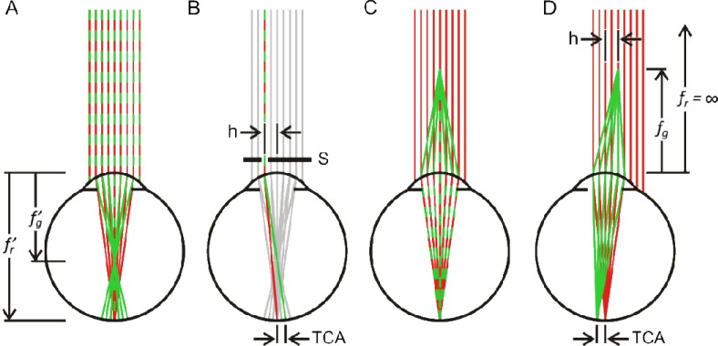
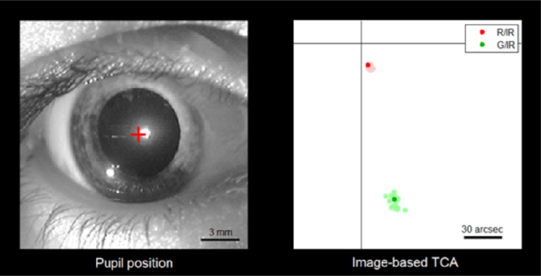
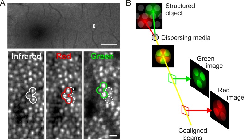
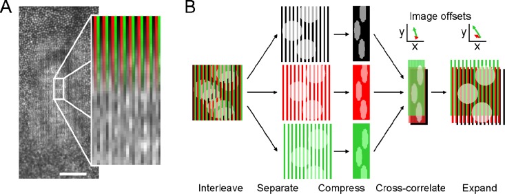
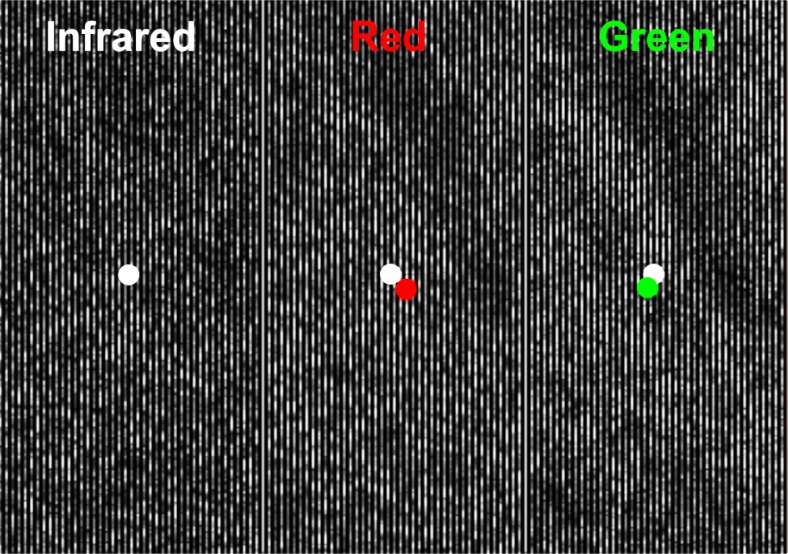
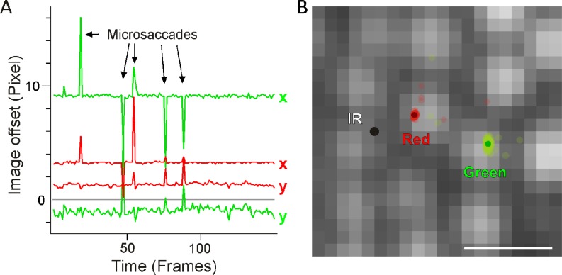
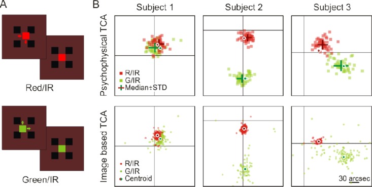
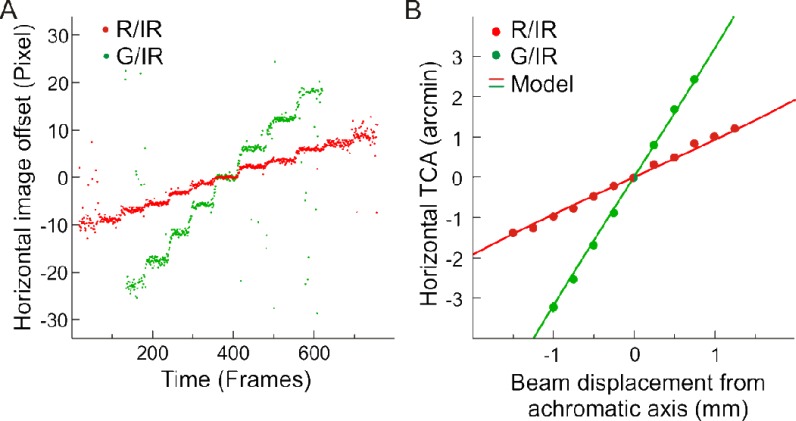
A special challenge arises when pursuing multi-wavelength imaging of retinal tissue in vivo, because the eye's optics must be used as the main focusing elements, and they introduce significant chromatic dispersion. Here we present an image-based method to measure and correct for the eye's transverse chromatic aberrations rapidly, non-invasively, and with high precision. We validate the technique against hyperacute psychophysical performance and the standard chromatic human eye model. In vivo correction of chromatic dispersion will enable confocal multi-wavelength images of the living retina to be aligned, and allow targeted chromatic stimulation of the photoreceptor mosaic to be performed accurately with sub-cellular resolution.
在进行视网膜组织的体内多波长成像时会出现一个特殊的挑战,因为眼睛的光学系统必须用作主要聚焦元件,而它们会引入显著的色散。在此,我们提出一种基于图像的方法,用于快速、非侵入性且高精度地测量和校正眼睛的横向色差。我们根据超急性心理物理学表现和标准的人眼色差模型对该技术进行了验证。体内色散校正将使活体视网膜的共焦多波长图像能够对齐,并允许以亚细胞分辨率精确地对光感受器镶嵌进行靶向色刺激。