Department of Prosthodontics, Ninth People's Hospital Affiliated to Shanghai Jiao Tong University, School of Medicine, Shanghai, China.
Int J Nanomedicine. 2013;8:257-65. doi: 10.2147/IJN.S39357. Epub 2013 Jan 11.
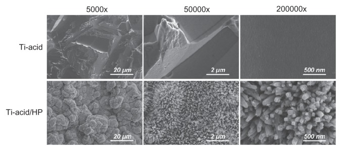
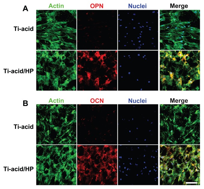
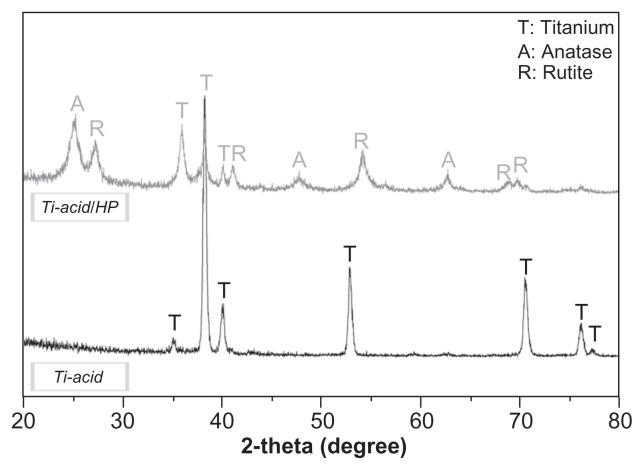
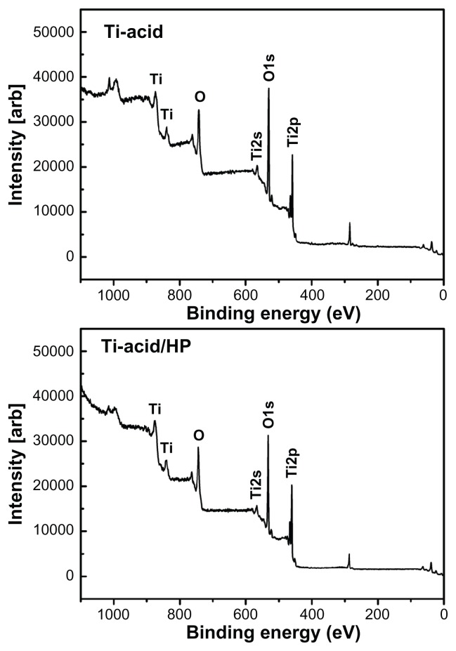
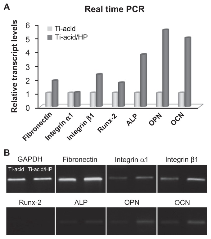
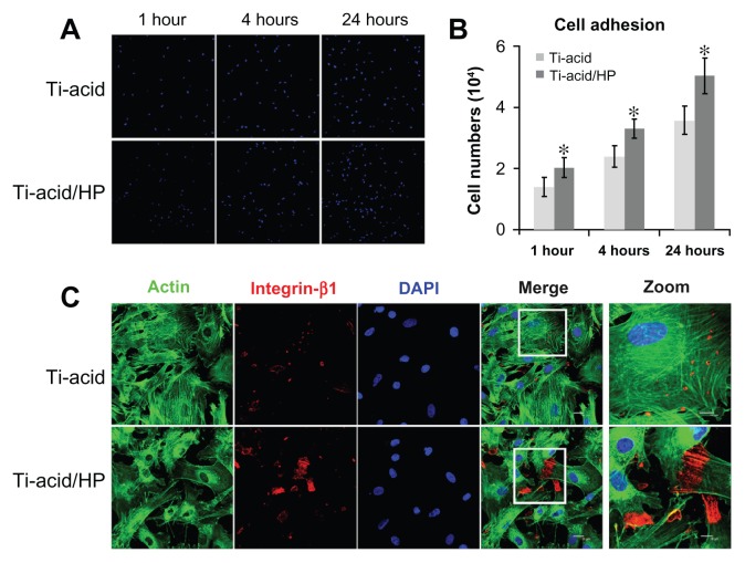
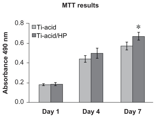
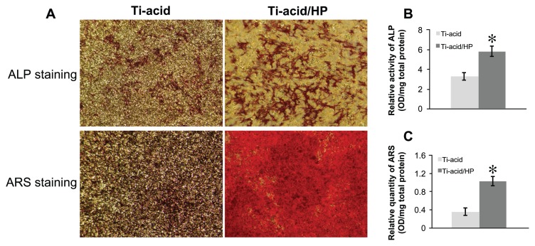
Various methods have been used to modify titanium implant surfaces with the aim of achieving better osseointegration. In this study, we fabricated a clustered nanorod structure on an acid-etched, microstructured titanium plate surface using hydrogen peroxide. We also evaluated biofunctionalization of the hybrid micro/nanorod topography on rat bone marrow mesenchymal stem cells. Scanning electron microscopy and x-ray diffraction were used to investigate the surface topography and phase composition of the modified titanium plate. Rat bone marrow mesenchymal stem cells were cultured and seeded on the plate. The adhesion ability of the cells was then assayed by cell counting at one, 4, and 24 hours after cell seeding, and expression of adhesion-related protein integrin β1 was detected by immunofluorescence. In addition, a polymerase chain reaction assay, alkaline phosphatase and Alizarin Red S staining assays, and osteopontin and osteocalcin immunofluorescence analyses were used to evaluate the osteogenic differentiation behavior of the cells.
The hybrid micro/nanoscale texture formed on the titanium surface enhanced the initial adhesion activity of the rat bone marrow mesenchymal stem cells. Importantly, the hierarchical structure promoted osteogenic differentiation of these cells.
This study suggests that a hybrid micro/nanorod topography on a titanium surface fabricated by treatment with hydrogen peroxide followed by acid etching might facilitate osseointegration of a titanium implant in vivo.
为了实现更好的骨整合,已经使用了各种方法来修饰钛植入物表面。在本研究中,我们使用过氧化氢在酸蚀、微结构化钛板表面上制造了集群纳米棒结构。我们还评估了混合微/纳米棒形貌对大鼠骨髓间充质干细胞的生物功能化作用。扫描电子显微镜和 X 射线衍射用于研究改性钛板的表面形貌和相组成。将大鼠骨髓间充质干细胞培养并接种在板上。在细胞接种后 1、4 和 24 小时,通过细胞计数测定细胞的粘附能力,并通过免疫荧光检测粘附相关蛋白整合素β1的表达。此外,还使用聚合酶链反应测定、碱性磷酸酶和茜素红 S 染色测定以及骨桥蛋白和骨钙素免疫荧光分析来评估细胞的成骨分化行为。
钛表面形成的混合微/纳米尺度结构增强了大鼠骨髓间充质干细胞的初始粘附活性。重要的是,分层结构促进了这些细胞的成骨分化。
本研究表明,通过过氧化氢处理后再进行酸蚀在钛表面上制造的混合微/纳米棒形貌可能有助于钛植入物在体内的骨整合。