• 文献检索
  • 文档翻译
  • 深度研究
  • 学术资讯
  • Suppr Zotero 插件Zotero 插件
  • 邀请有礼
  • 套餐&价格
  • 历史记录
应用&插件
Suppr Zotero 插件Zotero 插件浏览器插件Mac 客户端Windows 客户端微信小程序
定价
高级版会员购买积分包购买API积分包
服务
文献检索文档翻译深度研究API 文档MCP 服务
关于我们
关于 Suppr公司介绍联系我们用户协议隐私条款
关注我们

Suppr 超能文献

核心技术专利:CN118964589B侵权必究
粤ICP备2023148730 号-1Suppr @ 2026

文献检索

告别复杂PubMed语法,用中文像聊天一样搜索,搜遍4000万医学文献。AI智能推荐,让科研检索更轻松。

立即免费搜索

文件翻译

保留排版,准确专业,支持PDF/Word/PPT等文件格式,支持 12+语言互译。

免费翻译文档

深度研究

AI帮你快速写综述,25分钟生成高质量综述,智能提取关键信息,辅助科研写作。

立即免费体验

评估纳米颗粒在微载体细胞培养系统中的长期效应。

Assessment of long-term effects of nanoparticles in a microcarrier cell culture system.

机构信息

Center for Medical Research, Medical University of Graz, Graz, Austria.

出版信息

PLoS One. 2013;8(2):e56791. doi: 10.1371/journal.pone.0056791. Epub 2013 Feb 14.

DOI:10.1371/journal.pone.0056791
PMID:23457616
原文链接:https://pmc.ncbi.nlm.nih.gov/articles/PMC3573004/
Abstract

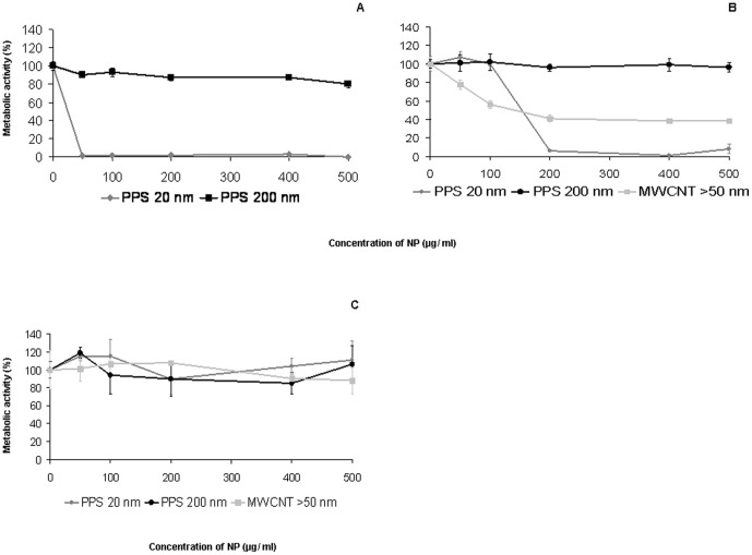
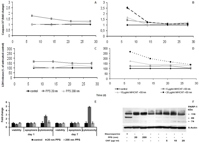
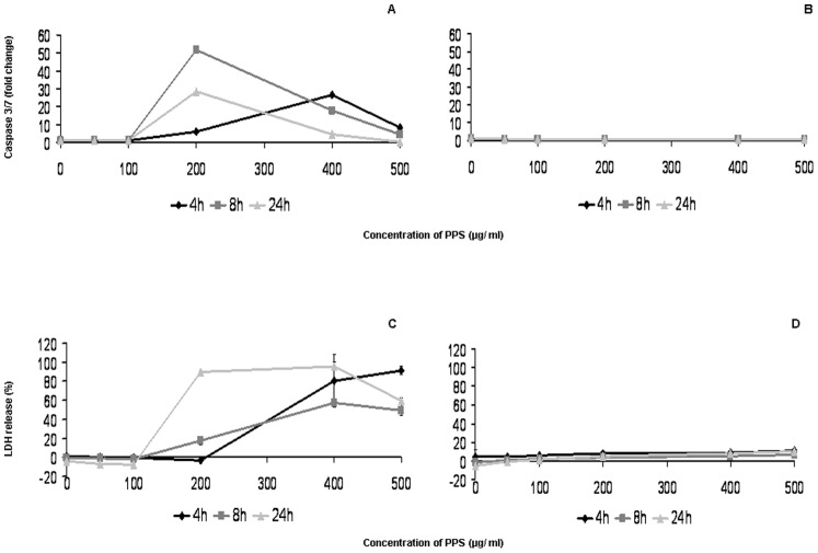
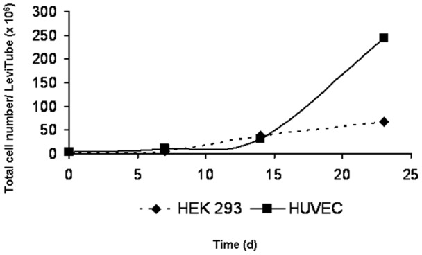
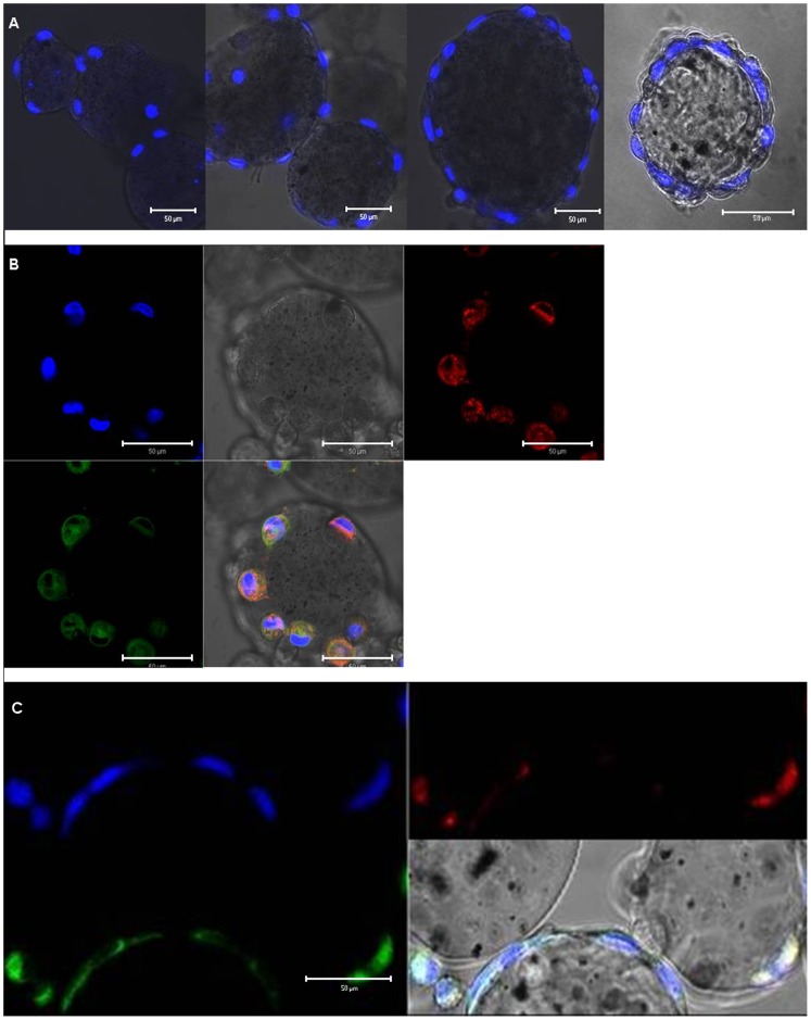
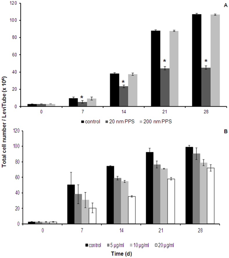
Nano-sized materials could find multiple applications in medical diagnosis and therapy. One main concern is that engineered nanoparticles, similar to combustion-derived nanoparticles, may cause adverse effects on human health by accumulation of entire particles or their degradation products. Chronic cytotoxicity must therefore be evaluated. In order to perform chronic cytotoxicity testing of plain polystyrene nanoparticles on the endothelial cell line EAhy 926, we established a microcarrier cell culture system for anchorage-dependent cells (BioLevitator(TM)). Cells were cultured for four weeks and exposed to doses, which were not cytotoxic upon 24 hours of exposure. For comparison, these particles were also studied in regularly sub-cultured cells, a method that has traditionally been used to assess chronic cellular effects. Culturing on basal membrane coated microcarriers produced very high cell densities. Fluorescent particles were mainly localized in the lysosomes of the exposed cells. After four weeks of exposure, the number of cells exposed to 20 nm polystyrene particles decreased by 60% as compared to untreated controls. When tested in sub-cultured cells, the same particles decreased cell numbers to 80% of the untreated controls. Dose-dependent decreases in cell numbers were also noted after exposure of microcarrier cultured cells to 50 nm short multi-walled carbon nanotubes. Our findings support that necrosis, but not apoptosis, contributed to cell death of the exposed cells in the microcarrier culture system. In conclusion, the established microcarrier model appears to be more sensitive for the identification of cellular effects upon prolonged and repeated exposure to nanoparticles than traditional sub-culturing.

摘要

纳米材料在医学诊断和治疗中有多种应用。一个主要的关注点是,工程纳米颗粒与燃烧衍生的纳米颗粒类似,可能会通过整个颗粒或其降解产物的积累对人体健康产生不良影响。因此,必须评估慢性细胞毒性。为了对内皮细胞系 EAhy 926 进行普通聚苯乙烯纳米颗粒的慢性细胞毒性测试,我们建立了一个锚定依赖性细胞(BioLevitator(TM))的微载体细胞培养系统。细胞培养了四周,并暴露于在 24 小时暴露时没有细胞毒性的剂量。为了进行比较,这些颗粒也在传统上用于评估慢性细胞效应的常规传代细胞中进行了研究。在基底膜包被的微载体上培养可产生非常高的细胞密度。荧光颗粒主要定位于暴露细胞的溶酶体中。与未处理的对照组相比,暴露于 20nm 聚苯乙烯颗粒的细胞数量在四周后减少了 60%。当在传代细胞中进行测试时,相同的颗粒将细胞数量减少到未处理对照组的 80%。暴露于微载体培养细胞的 50nm 短多壁碳纳米管后,还观察到细胞数量的剂量依赖性下降。我们的研究结果表明,在微载体培养系统中,暴露细胞的细胞死亡是由坏死引起的,而不是凋亡。总之,与传统的传代培养相比,建立的微载体模型似乎更能敏感地识别纳米颗粒长时间和重复暴露后的细胞效应。

https://cdn.ncbi.nlm.nih.gov/pmc/blobs/6577/3573004/b40e0e9dcf90/pone.0056791.g006.jpg
https://cdn.ncbi.nlm.nih.gov/pmc/blobs/6577/3573004/42a27340f9b6/pone.0056791.g001.jpg
https://cdn.ncbi.nlm.nih.gov/pmc/blobs/6577/3573004/800943fac52f/pone.0056791.g002.jpg
https://cdn.ncbi.nlm.nih.gov/pmc/blobs/6577/3573004/f3a6b7cce38d/pone.0056791.g003.jpg
https://cdn.ncbi.nlm.nih.gov/pmc/blobs/6577/3573004/c50f3aac9d9b/pone.0056791.g004.jpg
https://cdn.ncbi.nlm.nih.gov/pmc/blobs/6577/3573004/fafbd7153b65/pone.0056791.g005.jpg
https://cdn.ncbi.nlm.nih.gov/pmc/blobs/6577/3573004/b40e0e9dcf90/pone.0056791.g006.jpg

相似文献

1
Assessment of long-term effects of nanoparticles in a microcarrier cell culture system.评估纳米颗粒在微载体细胞培养系统中的长期效应。
PLoS One. 2013;8(2):e56791. doi: 10.1371/journal.pone.0056791. Epub 2013 Feb 14.
2
Comparison of two in vitro systems to assess cellular effects of nanoparticles-containing aerosols.比较两种体外系统评估含纳米颗粒气溶胶对细胞的影响。
Toxicol In Vitro. 2013 Feb;27(1):409-17. doi: 10.1016/j.tiv.2012.08.008. Epub 2012 Aug 10.
3
Cytotoxicity of nanoparticles independent from oxidative stress.纳米颗粒的细胞毒性与氧化应激无关。
J Toxicol Sci. 2009 Oct;34(4):363-75. doi: 10.2131/jts.34.363.
4
Cationic polystyrene nanosphere toxicity depends on cell-specific endocytic and mitochondrial injury pathways.阳离子聚苯乙烯纳米球的毒性取决于细胞特异性内吞和线粒体损伤途径。
ACS Nano. 2008 Jan;2(1):85-96. doi: 10.1021/nn700256c.
5
Assessing toxicity of fine and nanoparticles: comparing in vitro measurements to in vivo pulmonary toxicity profiles.评估细颗粒和纳米颗粒的毒性:将体外测量结果与体内肺部毒性概况进行比较。
Toxicol Sci. 2007 May;97(1):163-80. doi: 10.1093/toxsci/kfm018. Epub 2007 Feb 14.
6
Cytotoxity of nanoparticles is influenced by size, proliferation and embryonic origin of the cells used for testing.纳米颗粒的细胞毒性受用于测试的细胞的大小、增殖和胚胎起源的影响。
Nanotoxicology. 2012 Jun;6(4):424-39. doi: 10.3109/17435390.2011.586478. Epub 2011 May 31.
7
High content analysis provides mechanistic insights on the pathways of toxicity induced by amine-modified polystyrene nanoparticles.高内涵分析为胺改性聚苯乙烯纳米颗粒诱导的毒性途径提供了机制性见解。
PLoS One. 2014 Sep 19;9(9):e108025. doi: 10.1371/journal.pone.0108025. eCollection 2014.
8
Comparison of polystyrene and hydrogel microcarriers for optical imaging of adherent cells.聚苯乙烯和水凝胶微载体对贴壁细胞光学成像的比较。
J Biomed Opt. 2024 Jun;29(Suppl 2):S22708. doi: 10.1117/1.JBO.29.S2.S22708. Epub 2024 Jun 13.
9
Time-resolved characterization of the mechanisms of toxicity induced by silica and amino-modified polystyrene on alveolar-like macrophages.时间分辨法研究二氧化硅和氨基化聚苯乙烯诱导肺泡样巨噬细胞毒性的作用机制。
Arch Toxicol. 2020 Jan;94(1):173-186. doi: 10.1007/s00204-019-02604-5. Epub 2019 Nov 1.
10
Action of polystyrene nanoparticles of different sizes on lysosomal function and integrity.不同粒径聚苯乙烯纳米颗粒对溶酶体功能和完整性的作用。
Part Fibre Toxicol. 2012 Jul 12;9:26. doi: 10.1186/1743-8977-9-26.

引用本文的文献

1
Antioxidant Defense in Primary Murine Lung Cells following Short- and Long-Term Exposure to Plastic Particles.短期和长期暴露于塑料颗粒后原代小鼠肺细胞中的抗氧化防御
Antioxidants (Basel). 2023 Jan 18;12(2):227. doi: 10.3390/antiox12020227.
2
Dose-Dependent Cytotoxicity of Polypropylene Microplastics (PP-MPs) in Two Freshwater Fishes.聚丙烯微塑料(PP-MPs)对两种淡水鱼类的剂量依赖性细胞毒性。
Int J Mol Sci. 2022 Nov 10;23(22):13878. doi: 10.3390/ijms232213878.
3
The Effect of Chronic Exposure of Graphene Nanoplates on the Viability and Motility of A549 Cells.

本文引用的文献

1
Mechanisms of toxicity by carbon nanotubes.碳纳米管的毒性机制。
Toxicol Mech Methods. 2013 Mar;23(3):178-95. doi: 10.3109/15376516.2012.754534. Epub 2013 Jan 18.
2
Nanotoxicology and nanoparticle safety in biomedical designs.生物医学设计中的纳米毒理学和纳米颗粒安全性。
Int J Nanomedicine. 2011;6:1117-27. doi: 10.2147/IJN.S16603. Epub 2011 May 31.
3
A comparison of acute and long-term effects of industrial multiwalled carbon nanotubes on human lung and immune cells in vitro.工业用多壁碳纳米管对体外人肺和免疫细胞的急性和长期效应比较。
石墨烯纳米片长期暴露对A549细胞活力和运动性的影响
Nanomaterials (Basel). 2022 Jun 16;12(12):2074. doi: 10.3390/nano12122074.
4
Screening for Effects of Inhaled Nanoparticles in Cell Culture Models for Prolonged Exposure.在细胞培养模型中对吸入性纳米颗粒进行长期暴露影响的筛选。
Nanomaterials (Basel). 2021 Feb 28;11(3):606. doi: 10.3390/nano11030606.
5
Potential and recent advances of microcarriers in repairing cartilage defects.微载体在修复软骨缺损方面的潜力及最新进展。
J Orthop Translat. 2021 Jan 13;27:101-109. doi: 10.1016/j.jot.2020.10.005. eCollection 2021 Mar.
6
New Insights into the Cell Death Signaling Pathways Triggered by Long-Term Exposure to Silicon-Based Quantum Dots in Human Lung Fibroblasts.长期暴露于硅基量子点的人肺成纤维细胞触发的细胞死亡信号通路新见解
Nanomaterials (Basel). 2021 Jan 27;11(2):323. doi: 10.3390/nano11020323.
7
Evaluation of the Hemocompatibility and Anticancer Potential of Poly(-Caprolactone) and Poly(3-Hydroxybutyrate) Microcarriers with Encapsulated Chrysin.聚(ε-己内酯)和聚(3-羟基丁酸酯)微载体包封白杨素的血液相容性和抗癌潜力评估
Pharmaceutics. 2021 Jan 16;13(1):109. doi: 10.3390/pharmaceutics13010109.
8
Proposing Urothelial and Muscle In Vitro Cell Models as a Novel Approach for Assessment of Long-Term Toxicity of Nanoparticles.提出尿路上皮和肌肉体外细胞模型作为评估纳米颗粒长期毒性的新方法。
Int J Mol Sci. 2020 Oct 13;21(20):7545. doi: 10.3390/ijms21207545.
9
p53 at the Crossroads between Different Types of HDAC Inhibitor-Mediated Cancer Cell Death.p53 在不同类型的 HDAC 抑制剂介导的癌细胞死亡中的作用。
Int J Mol Sci. 2019 May 15;20(10):2415. doi: 10.3390/ijms20102415.
10
Tissue Regeneration of Human Mesenchymal Stem Cells on Porous Gelatin Micro-Carriers by Long-Term Dynamic Culture.多孔明胶微载体长期动态培养对人骨髓间充质干细胞的组织再生。
Tissue Eng Regen Med. 2019 Jan 28;16(1):19-28. doi: 10.1007/s13770-018-00174-8. eCollection 2019 Feb.
Toxicol Lett. 2011 Feb 5;200(3):176-86. doi: 10.1016/j.toxlet.2010.11.012. Epub 2010 Nov 26.
4
Bioaccumulation and toxicity of gold nanoparticles after repeated administration in mice.金纳米粒子在小鼠体内重复给药后的生物积累和毒性。
Biochem Biophys Res Commun. 2010 Mar 19;393(4):649-55. doi: 10.1016/j.bbrc.2010.02.046. Epub 2010 Feb 12.
5
Biodistribution of gold nanoparticles in mouse lung following intratracheal instillation.气管内滴注后金纳米颗粒在小鼠肺内的生物分布。
Chem Cent J. 2009 Nov 20;3:16. doi: 10.1186/1752-153X-3-16.
6
Assessment of the dermal and ocular irritation potential of multi-walled carbon nanotubes by using in vitro and in vivo methods.运用体外和体内方法评估多壁碳纳米管的皮肤和眼刺激潜力。
Toxicol Lett. 2009 Dec 15;191(2-3):268-74. doi: 10.1016/j.toxlet.2009.09.007. Epub 2009 Sep 19.
7
Nanoparticles, human health hazard and regulation.纳米颗粒、人体健康危害与监管
J R Soc Interface. 2010 Feb 6;7 Suppl 1(Suppl 1):S119-29. doi: 10.1098/rsif.2009.0252.focus. Epub 2009 Sep 2.
8
Inhaled fluorescent magnetic nanoparticles induced extramedullary hematopoiesis in the spleen of mice.吸入式荧光磁性纳米颗粒可诱导小鼠脾脏产生髓外造血。
J Occup Health. 2009;51(5):423-31. doi: 10.1539/joh.l8159. Epub 2009 Aug 25.
9
Cytotoxicity of nanoparticles independent from oxidative stress.纳米颗粒的细胞毒性与氧化应激无关。
J Toxicol Sci. 2009 Oct;34(4):363-75. doi: 10.2131/jts.34.363.
10
Toxicity and penetration of TiO2 nanoparticles in hairless mice and porcine skin after subchronic dermal exposure.二氧化钛纳米颗粒在亚慢性皮肤暴露后对无毛小鼠和猪皮肤的毒性及渗透情况
Toxicol Lett. 2009 Dec 1;191(1):1-8. doi: 10.1016/j.toxlet.2009.05.020. Epub 2009 Jun 6.