Oh Jaeryung, Yoo Chungkwon, Yun Cheol Min, Yang Kyung-Sook, Kim Seong-Woo, Huh Kuhl
Department of Ophthalmology, Korea University College of Medicine, Seoul, Korea.
Korean J Ophthalmol. 2013 Jun;27(3):172-7. doi: 10.3341/kjo.2013.27.3.172. Epub 2013 Apr 30.
To evaluate a simplified method to measure peripapillary choroidal thickness using commercially available, three-dimensional optical coherence tomography (3D-OCT).
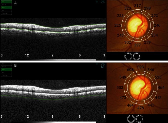
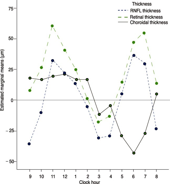
3D-OCT images of normal eyes were consecutively obtained from the 3D-OCT database of Korea University Medical Center On the peripapillary images for retinal nerve fiber layer (RNFL) analysis, choroidal thickness was measured by adjusting the segmentation line for the retinal pigment epithelium to the chorioscleral junction using the modification tool built into the 3D-OCT image viewer program. Variations of choroidal thickness at 12 sectors of the peripapillary area were evaluated.
We were able to measure the peripapillary choroidal thickness in 40 eyes of our 40 participants, who had a mean age of 41.2 years (range, 15 to 84 years). Choroidal thickness measurements had strong inter-observer correlation at each sector (r = 0.901 to 0.991, p < 0.001). The mean choroidal thickness was 191 ± 62 µm. Choroidal thickness was greatest at the temporal quadrant (mean ± SD, 210 ± 78 µm), followed by the superior (202 ± 66 µm), nasal (187 ± 64 µm), and inferior quadrants (152 ± 59 µm).
The measurement of choroidal thickness on peripapillary circle scan images for RNFL analysis using the 3D-OCT viewing program was highly reliable and efficient.
评估一种使用商用三维光学相干断层扫描(3D-OCT)测量视乳头周围脉络膜厚度的简化方法。
从韩国大学医学中心的3D-OCT数据库中连续获取正常眼睛的3D-OCT图像。在用于视网膜神经纤维层(RNFL)分析的视乳头周围图像上,使用3D-OCT图像查看程序中内置的修改工具,将视网膜色素上皮的分割线调整到脉络膜巩膜交界处来测量脉络膜厚度。评估视乳头周围区域12个扇区的脉络膜厚度变化。
我们能够在40名参与者的40只眼中测量视乳头周围脉络膜厚度,这些参与者的平均年龄为41.2岁(范围为15至84岁)。各扇区的脉络膜厚度测量在观察者间具有很强的相关性(r = 0.901至0.991,p < 0.001)。平均脉络膜厚度为191±62μm。脉络膜厚度在颞侧象限最大(平均值±标准差,210±78μm),其次是上方(202±66μm)、鼻侧(187±64μm)和下方象限(152±59μm)。
使用3D-OCT查看程序在用于RNFL分析的视乳头周围环形扫描图像上测量脉络膜厚度是高度可靠且有效的。