• 文献检索
  • 文档翻译
  • 深度研究
  • 学术资讯
  • Suppr Zotero 插件Zotero 插件
  • 邀请有礼
  • 套餐&价格
  • 历史记录
应用&插件
Suppr Zotero 插件Zotero 插件浏览器插件Mac 客户端Windows 客户端微信小程序
定价
高级版会员购买积分包购买API积分包
服务
文献检索文档翻译深度研究API 文档MCP 服务
关于我们
关于 Suppr公司介绍联系我们用户协议隐私条款
关注我们

Suppr 超能文献

核心技术专利:CN118964589B侵权必究
粤ICP备2023148730 号-1Suppr @ 2026

文献检索

告别复杂PubMed语法,用中文像聊天一样搜索,搜遍4000万医学文献。AI智能推荐,让科研检索更轻松。

立即免费搜索

文件翻译

保留排版,准确专业,支持PDF/Word/PPT等文件格式,支持 12+语言互译。

免费翻译文档

深度研究

AI帮你快速写综述,25分钟生成高质量综述,智能提取关键信息,辅助科研写作。

立即免费体验

多个染色组织学图像的三维重建

3D reconstruction of multiple stained histology images.

作者信息

Song Yi, Treanor Darren, Bulpitt Andrew J, Magee Derek R

机构信息

School of Computing, University of Leeds, Leeds, UK.

出版信息

J Pathol Inform. 2013 Mar 30;4(Suppl):S7. doi: 10.4103/2153-3539.109864. Print 2013.

DOI:10.4103/2153-3539.109864
PMID:23766943
原文链接:https://pmc.ncbi.nlm.nih.gov/articles/PMC3678754/
Abstract

CONTEXT

Three dimensional (3D) tissue reconstructions from the histology images with different stains allows the spatial alignment of structural and functional elements highlighted by different stains for quantitative study of many physiological and pathological phenomena. This has significant potential to improve the understanding of the growth patterns and the spatial arrangement of diseased cells, and enhance the study of biomechanical behavior of the tissue structures towards better treatments (e.g. tissue-engineering applications).

METHODS

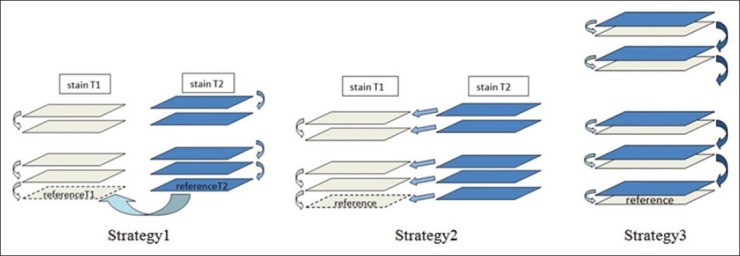
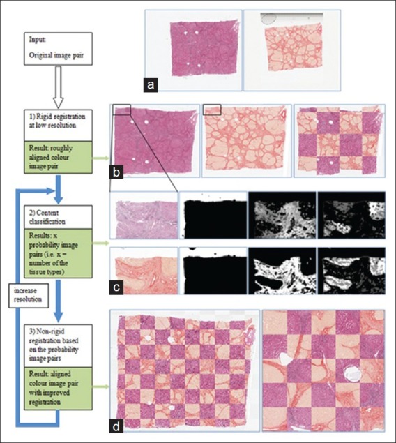
This paper evaluates three strategies for 3D reconstruction from sets of two dimensional (2D) histological sections with different stains, by combining methods of 2D multi-stain registration and 3D volumetric reconstruction from same stain sections.

SETTING AND DESIGN

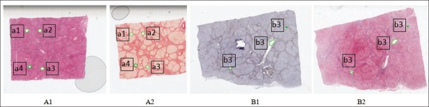
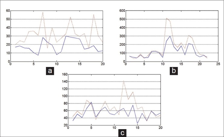
The different strategies have been evaluated on two liver specimens (80 sections in total) stained with Hematoxylin and Eosin (H and E), Sirius Red, and Cytokeratin (CK) 7.

RESULTS AND CONCLUSION

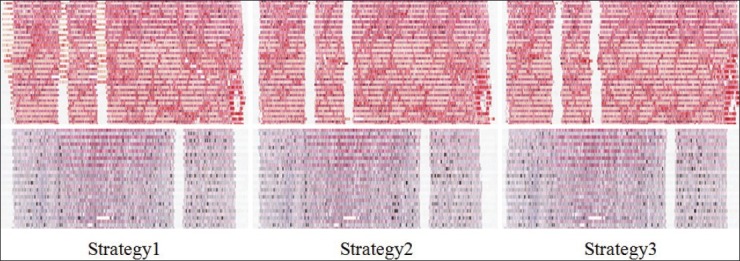
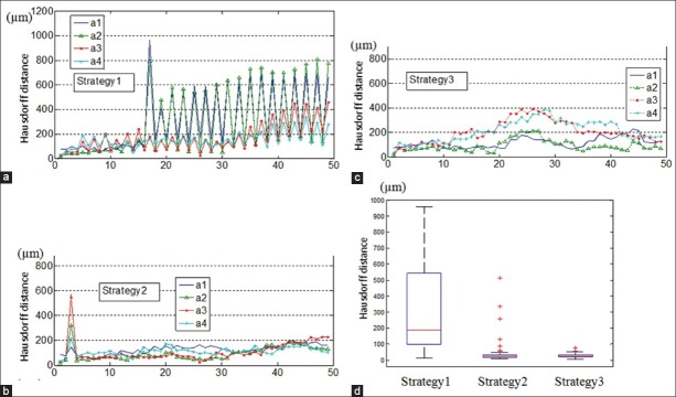
A strategy of using multi-stain registration to align images of a second stain to a volume reconstructed by same-stain registration results in the lowest overall error, although an interlaced image registration approach may be more robust to poor section quality.

摘要

背景

利用不同染色的组织学图像进行三维(3D)组织重建,能够使不同染色突出显示的结构和功能元素在空间上对齐,从而对多种生理和病理现象进行定量研究。这对于增进对病变细胞生长模式和空间排列的理解,以及加强对组织结构生物力学行为的研究以实现更好的治疗(如组织工程应用)具有巨大潜力。

方法

本文通过结合二维多染色配准方法和来自相同染色切片的三维体积重建方法,评估了从具有不同染色的二维(2D)组织学切片集进行三维重建的三种策略。

设置与设计

在两个用苏木精和伊红(H&E)、天狼星红以及细胞角蛋白(CK)7染色的肝脏标本(总共80个切片)上评估了不同策略。

结果与结论

使用多染色配准将第二种染色图像与通过相同染色配准重建的体积对齐的策略总体误差最低,尽管隔行图像配准方法可能对较差的切片质量更具鲁棒性。

https://cdn.ncbi.nlm.nih.gov/pmc/blobs/204f/3678754/fce00fc0d88b/JPI-4-7-g007.jpg
https://cdn.ncbi.nlm.nih.gov/pmc/blobs/204f/3678754/9fac11cffed8/JPI-4-7-g001.jpg
https://cdn.ncbi.nlm.nih.gov/pmc/blobs/204f/3678754/0d299fc4476d/JPI-4-7-g002.jpg
https://cdn.ncbi.nlm.nih.gov/pmc/blobs/204f/3678754/9ae789712f0b/JPI-4-7-g003.jpg
https://cdn.ncbi.nlm.nih.gov/pmc/blobs/204f/3678754/feb069cb57af/JPI-4-7-g005.jpg
https://cdn.ncbi.nlm.nih.gov/pmc/blobs/204f/3678754/1339687fd80e/JPI-4-7-g006.jpg
https://cdn.ncbi.nlm.nih.gov/pmc/blobs/204f/3678754/fce00fc0d88b/JPI-4-7-g007.jpg

相似文献

1
3D reconstruction of multiple stained histology images.多个染色组织学图像的三维重建
J Pathol Inform. 2013 Mar 30;4(Suppl):S7. doi: 10.4103/2153-3539.109864. Print 2013.
2
Reconstruction of 3-dimensional histology volume and its application to study mouse mammary glands.三维组织学体积重建及其在小鼠乳腺研究中的应用。
J Vis Exp. 2014 Jul 26(89):e51325. doi: 10.3791/51325.
3
Robust 3D image reconstruction of pancreatic cancer tumors from histopathological images with different stains and its quantitative performance evaluation.利用不同染色的组织病理学图像对胰腺癌肿瘤进行稳健的三维图像重建及其定量性能评估。
Int J Comput Assist Radiol Surg. 2019 Dec;14(12):2047-2055. doi: 10.1007/s11548-019-02019-8. Epub 2019 Jul 2.
4
Robust joint registration of multiple stains and MRI for multimodal 3D histology reconstruction: Application to the Allen human brain atlas.多模态 3D 组织学重建的多染色和 MRI 的稳健联合配准:Allen 人类大脑图谱的应用。
Med Image Anal. 2022 Jan;75:102265. doi: 10.1016/j.media.2021.102265. Epub 2021 Oct 16.
5
2D and 3D MALDI-imaging: conceptual strategies for visualization and data mining.二维和三维基质辅助激光解吸电离成像:可视化与数据挖掘的概念策略
Biochim Biophys Acta. 2014 Jan;1844(1 Pt A):117-37. doi: 10.1016/j.bbapap.2013.01.040. Epub 2013 Mar 4.
6
Deep learning based registration of serial whole-slide histopathology images in different stains.基于深度学习的不同染色的连续全切片组织病理学图像配准
J Pathol Inform. 2023 Apr 23;14:100311. doi: 10.1016/j.jpi.2023.100311. eCollection 2023.
7
Accurate and Robust Alignment of Differently Stained Histologic Images Based on Greedy Diffeomorphic Registration.基于贪婪微分同胚配准的不同染色组织学图像的精确且稳健配准
Appl Sci (Basel). 2021 Feb;11(4). doi: 10.3390/app11041892. Epub 2021 Feb 21.
8
Unsupervised content classification based nonrigid registration of differently stained histology images.基于无监督内容分类的不同染色组织学图像非刚性配准
IEEE Trans Biomed Eng. 2014 Jan;61(1):96-108. doi: 10.1109/TBME.2013.2277777. Epub 2013 Aug 8.
9
Three-dimensional histopathological reconstruction of bladder tumours.膀胱肿瘤的三维组织病理学重建
Diagn Pathol. 2019 Mar 28;14(1):25. doi: 10.1186/s13000-019-0803-7.
10
A Survey of Methods for 3D Histology Reconstruction.三维组织学重建方法研究综述。
Med Image Anal. 2018 May;46:73-105. doi: 10.1016/j.media.2018.02.004. Epub 2018 Feb 21.

引用本文的文献

1
Serial section histopathology image dataset of colon and pancreas tissue captured through light microscope.通过光学显微镜拍摄的结肠和胰腺组织连续切片组织病理学图像数据集。
Data Brief. 2024 Apr 15;54:110429. doi: 10.1016/j.dib.2024.110429. eCollection 2024 Jun.
2
A Multi-Stain Breast Cancer Histological Whole-Slide-Image Data Set from Routine Diagnostics.多染色乳腺癌组织学全切片图像数据集来自常规诊断。
Sci Data. 2023 Aug 24;10(1):562. doi: 10.1038/s41597-023-02422-6.
3
Virtual alignment of pathology image series for multi-gigapixel whole slide images.

本文引用的文献

1
Toward routine use of 3D histopathology as a research tool.迈向将 3D 组织病理学常规用作研究工具。
Am J Pathol. 2012 May;180(5):1835-42. doi: 10.1016/j.ajpath.2012.01.033. Epub 2012 Apr 9.
2
Real-time deformable registration of multi-modal whole slides for digital pathology.多模态全切片的数字病理学实时可变形配准。
Comput Med Imaging Graph. 2011 Oct-Dec;35(7-8):542-56. doi: 10.1016/j.compmedimag.2011.06.006. Epub 2011 Jun 28.
3
Position, rotation, and scale invariant optical correlation.位置、旋转和尺度不变光学相关
多吉帕像素全玻片图像的病理学图像系列的虚拟配准。
Nat Commun. 2023 Jul 26;14(1):4502. doi: 10.1038/s41467-023-40218-9.
4
Generating post-hoc explanation from deep neural networks for multi-modal medical image analysis tasks.从深度神经网络生成事后解释以用于多模态医学图像分析任务。
MethodsX. 2023 Jan 10;10:102009. doi: 10.1016/j.mex.2023.102009. eCollection 2023.
5
Tissue clearing and 3D reconstruction of digitized, serially sectioned slides provide novel insights into pancreatic cancer.数字化、连续切片的组织透明化和 3D 重建为胰腺癌研究提供了新的视角。
Med. 2023 Feb 10;4(2):75-91. doi: 10.1016/j.medj.2022.11.009. Epub 2023 Jan 24.
6
CODA: quantitative 3D reconstruction of large tissues at cellular resolution.CODA:用于细胞分辨率的大型组织的定量 3D 重建。
Nat Methods. 2022 Nov;19(11):1490-1499. doi: 10.1038/s41592-022-01650-9. Epub 2022 Oct 24.
7
Robust joint registration of multiple stains and MRI for multimodal 3D histology reconstruction: Application to the Allen human brain atlas.多模态 3D 组织学重建的多染色和 MRI 的稳健联合配准:Allen 人类大脑图谱的应用。
Med Image Anal. 2022 Jan;75:102265. doi: 10.1016/j.media.2021.102265. Epub 2021 Oct 16.
8
Tutorial: methods for three-dimensional visualization of archival tissue material.教程:存档组织材料的三维可视化方法
Nat Protoc. 2021 Nov;16(11):4945-4962. doi: 10.1038/s41596-021-00611-4. Epub 2021 Oct 29.
9
Multimodal 3D photoacoustic remote sensing and confocal fluorescence microscopy imaging.多模态 3D 光声远程传感与共聚焦荧光显微镜成像。
J Biomed Opt. 2021 Sep;26(9). doi: 10.1117/1.JBO.26.9.096501.
10
Improving Algorithm for the Alignment of Consecutive, Whole-Slide, Immunohistochemical Section Images.用于连续全切片免疫组化切片图像配准的改进算法
J Pathol Inform. 2021 Aug 3;12:29. doi: 10.4103/jpi.jpi_106_20. eCollection 2021.
Appl Opt. 1976 Jul 1;15(7):1795-9. doi: 10.1364/AO.15.001795.
4
Feature-based registration of histopathology images with different stains: an application for computerized follicular lymphoma prognosis.不同染色的组织病理学图像基于特征的配准:在计算机化滤泡性淋巴瘤预后评估中的应用
Comput Methods Programs Biomed. 2009 Dec;96(3):182-92. doi: 10.1016/j.cmpb.2009.04.012. Epub 2009 May 31.
5
Three dimensional reconstruction of a human breast carcinoma using routine laboratory equipment and immunohistochemistry.使用常规实验室设备和免疫组织化学对人乳腺癌进行三维重建。
J Clin Pathol. 2005 Sep;58(9):968-72. doi: 10.1136/jcp.2004.024794.
6
Piecewise affine registration of biological images for volume reconstruction.用于体积重建的生物图像分段仿射配准
Med Image Anal. 2006 Jun;10(3):465-83. doi: 10.1016/j.media.2005.03.008. Epub 2005 Jun 15.