• 文献检索
  • 文档翻译
  • 深度研究
  • 学术资讯
  • Suppr Zotero 插件Zotero 插件
  • 邀请有礼
  • 套餐&价格
  • 历史记录
应用&插件
Suppr Zotero 插件Zotero 插件浏览器插件Mac 客户端Windows 客户端微信小程序
定价
高级版会员购买积分包购买API积分包
服务
文献检索文档翻译深度研究API 文档MCP 服务
关于我们
关于 Suppr公司介绍联系我们用户协议隐私条款
关注我们

Suppr 超能文献

核心技术专利:CN118964589B侵权必究
粤ICP备2023148730 号-1Suppr @ 2026

文献检索

告别复杂PubMed语法,用中文像聊天一样搜索,搜遍4000万医学文献。AI智能推荐,让科研检索更轻松。

立即免费搜索

文件翻译

保留排版,准确专业,支持PDF/Word/PPT等文件格式,支持 12+语言互译。

免费翻译文档

深度研究

AI帮你快速写综述,25分钟生成高质量综述,智能提取关键信息,辅助科研写作。

立即免费体验

基于图谱的头颈部危及器官和淋巴结靶区自动勾画:一项临床验证。

Atlas-based automatic segmentation of head and neck organs at risk and nodal target volumes: a clinical validation.

机构信息

Radiation Oncology Dept, Clinique & Maternité Ste-Elisabeth, Place Louise Godin 15, 5000 - Namur, Belgium.

出版信息

Radiat Oncol. 2013 Jun 26;8:154. doi: 10.1186/1748-717X-8-154.

DOI:10.1186/1748-717X-8-154
PMID:23803232
原文链接:https://pmc.ncbi.nlm.nih.gov/articles/PMC3722083/
Abstract

BACKGROUND

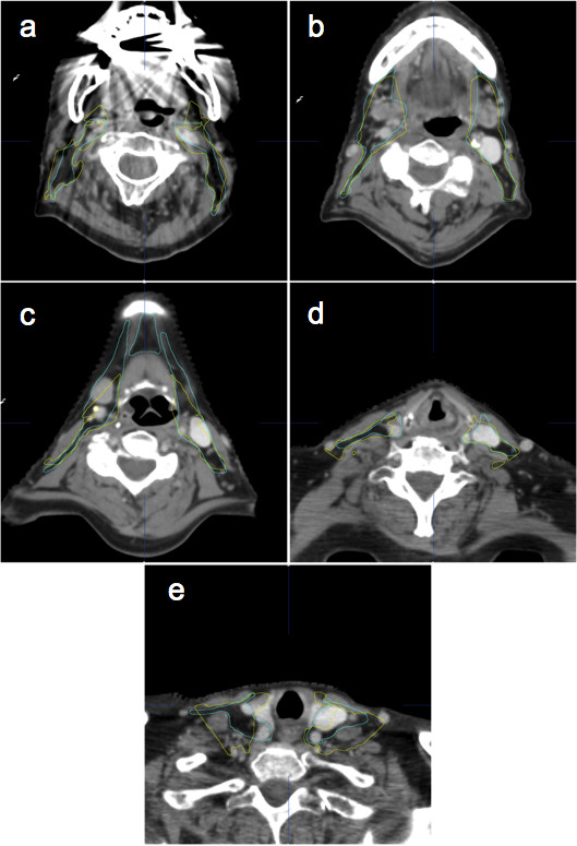
Intensity modulated radiotherapy for head and neck cancer necessitates accurate definition of organs at risk (OAR) and clinical target volumes (CTV). This crucial step is time consuming and prone to inter- and intra-observer variations. Automatic segmentation by atlas deformable registration may help to reduce time and variations. We aim to test a new commercial atlas algorithm for automatic segmentation of OAR and CTV in both ideal and clinical conditions.

METHODS

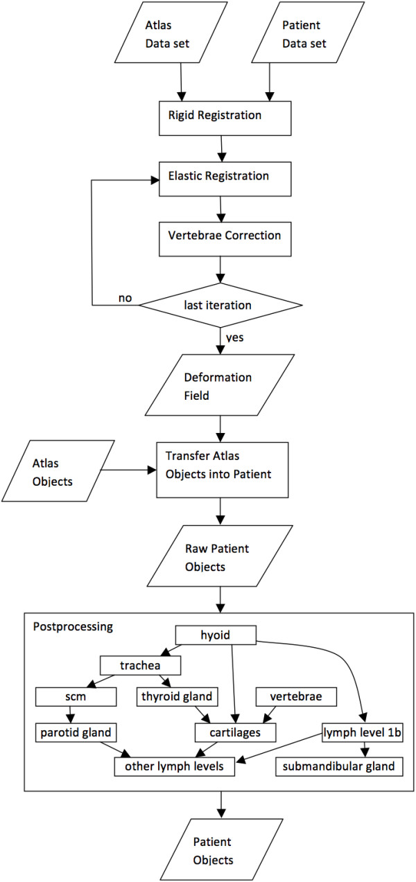
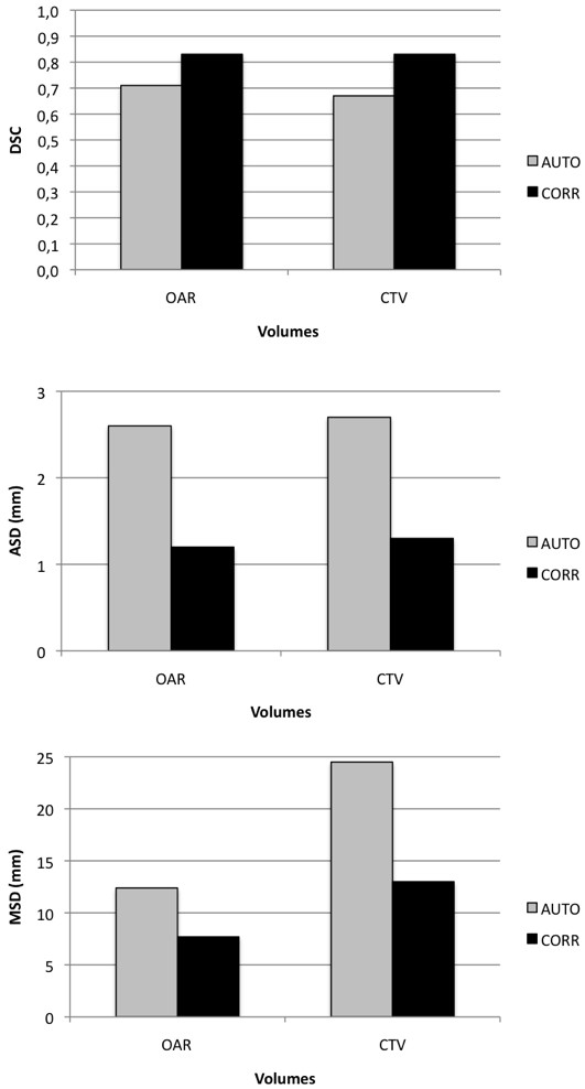
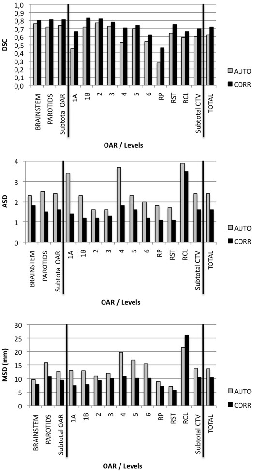
The updated Brainlab automatic head and neck atlas segmentation was tested on 20 patients: 10 cN0-stages (ideal population) and 10 unselected N-stages (clinical population). Following manual delineation of OAR and CTV, automatic segmentation of the same set of structures was performed and afterwards manually corrected. Dice Similarity Coefficient (DSC), Average Surface Distance (ASD) and Maximal Surface Distance (MSD) were calculated for "manual to automatic" and "manual to corrected" volumes comparisons.

RESULTS

In both groups, automatic segmentation saved about 40% of the corresponding manual segmentation time. This effect was more pronounced for OAR than for CTV. The edition of the automatically obtained contours significantly improved DSC, ASD and MSD. Large distortions of normal anatomy or lack of iodine contrast were the limiting factors.

CONCLUSIONS

The updated Brainlab atlas-based automatic segmentation tool for head and neck Cancer patients is timesaving but still necessitates review and corrections by an expert.

摘要

背景

头颈部癌症的强度调制放疗需要准确定义危及器官(OAR)和临床靶区(CTV)。这一关键步骤既耗时又容易出现观察者间和观察者内的差异。基于图谱的变形配准的自动分割可能有助于减少时间和差异。我们旨在测试一种新的商业图谱算法,以在理想和临床条件下自动分割 OAR 和 CTV。

方法

对 20 名患者(10 名 cN0 期(理想人群)和 10 名未选择的 N 期(临床人群))进行了更新的 Brainlab 自动头颈部图谱分割测试。在手动勾画 OAR 和 CTV 之后,对同一组结构进行了自动分割,然后进行手动校正。计算了“手动到自动”和“手动到校正”体积比较的 Dice 相似性系数(DSC)、平均表面距离(ASD)和最大表面距离(MSD)。

结果

在两组中,自动分割都节省了约 40%的相应手动分割时间。OAR 的效果比 CTV 更明显。自动获得的轮廓的编辑显著提高了 DSC、ASD 和 MSD。正常解剖结构的大变形或缺乏碘对比是限制因素。

结论

用于头颈部癌症患者的更新的基于 Brainlab 图谱的自动分割工具可以节省时间,但仍需要专家进行审查和校正。

https://cdn.ncbi.nlm.nih.gov/pmc/blobs/dba0/3722083/44b06647c310/1748-717X-8-154-5.jpg
https://cdn.ncbi.nlm.nih.gov/pmc/blobs/dba0/3722083/088af7e97992/1748-717X-8-154-1.jpg
https://cdn.ncbi.nlm.nih.gov/pmc/blobs/dba0/3722083/6e90e45cad66/1748-717X-8-154-2.jpg
https://cdn.ncbi.nlm.nih.gov/pmc/blobs/dba0/3722083/5ac4f3ee4b12/1748-717X-8-154-3.jpg
https://cdn.ncbi.nlm.nih.gov/pmc/blobs/dba0/3722083/7c6a962b8fa9/1748-717X-8-154-4.jpg
https://cdn.ncbi.nlm.nih.gov/pmc/blobs/dba0/3722083/44b06647c310/1748-717X-8-154-5.jpg

相似文献

1
Atlas-based automatic segmentation of head and neck organs at risk and nodal target volumes: a clinical validation.基于图谱的头颈部危及器官和淋巴结靶区自动勾画:一项临床验证。
Radiat Oncol. 2013 Jun 26;8:154. doi: 10.1186/1748-717X-8-154.
2
Automatic segmentation of head and neck CT images for radiotherapy treatment planning using multiple atlases, statistical appearance models, and geodesic active contours.使用多个图谱、统计外观模型和测地线活动轮廓对头部和颈部CT图像进行自动分割以用于放射治疗计划
Med Phys. 2014 May;41(5):051910. doi: 10.1118/1.4871623.
3
Clinical evaluation of multi-atlas based segmentation of lymph node regions in head and neck and prostate cancer patients.基于多图谱的头颈部和前列腺癌患者淋巴结区域分割的临床评估。
Radiat Oncol. 2013 Oct 3;8:229. doi: 10.1186/1748-717X-8-229.
4
Geometric and dosimetric evaluations of atlas-based segmentation methods of MR images in the head and neck region.基于图谱的头部和颈部磁共振图像分割方法的几何和剂量学评估。
Phys Med Biol. 2018 Jul 11;63(14):145007. doi: 10.1088/1361-6560/aacb65.
5
The feasibility of atlas-based automatic segmentation of MRI for H&N radiotherapy planning.基于图谱的MRI自动分割在头颈部放疗计划中的可行性。
J Appl Clin Med Phys. 2016 Jul 8;17(4):146-154. doi: 10.1120/jacmp.v17i4.6051.
6
Validation of clinical acceptability of an atlas-based segmentation algorithm for the delineation of organs at risk in head and neck cancer.基于图谱的分割算法用于头颈癌中危及器官 delineation 的临床可接受性验证。 (注:“delineation”这个词可能有误,正确的或许是“delineation”,可结合具体语境进一步确认准确含义并调整译文)
Med Phys. 2015 Sep;42(9):5027-34. doi: 10.1118/1.4927567.
7
Comparison of atlas-based and deep learning methods for organs at risk delineation on head-and-neck CT images using an automated treatment planning system.使用自动治疗计划系统,对头颈部CT图像上的危及器官进行勾画时,基于图谱的方法与深度学习方法的比较。
Radiother Oncol. 2022 Dec;177:61-70. doi: 10.1016/j.radonc.2022.10.029. Epub 2022 Nov 1.
8
Improving automatic delineation for head and neck organs at risk by Deep Learning Contouring.深度学习勾画技术对头颈部危险器官自动勾画的改善。
Radiother Oncol. 2020 Jan;142:115-123. doi: 10.1016/j.radonc.2019.09.022. Epub 2019 Oct 22.
9
Evaluation of auto-segmentation accuracy of cloud-based artificial intelligence and atlas-based models.基于云的人工智能和图谱模型的自动分割准确性评估。
Radiat Oncol. 2021 Sep 9;16(1):175. doi: 10.1186/s13014-021-01896-1.
10
Evaluation of automatic atlas-based lymph node segmentation for head-and-neck cancer.头颈部癌症自动图谱基于淋巴结分割的评估。
Int J Radiat Oncol Biol Phys. 2010 Jul 1;77(3):959-66. doi: 10.1016/j.ijrobp.2009.09.023. Epub 2010 Mar 16.

引用本文的文献

1
Comparative Analysis of Atlas and Neural Network Autosegmentation Methods for Pediatric Craniospinal Irradiation With the Development of a Knowledge-Based Quality Assurance Tool.基于知识的质量保证工具开发下小儿颅脊髓照射图谱与神经网络自动分割方法的比较分析
Adv Radiat Oncol. 2025 Jul 28;10(9):101847. doi: 10.1016/j.adro.2025.101847. eCollection 2025 Sep.
2
Domain-Adaptive and Per-Fraction Guided Deep Learning Framework for Magnetic Resonance Imaging-Based Segmentation of Organs at Risk in Gynecologic Cancers.用于基于磁共振成像的妇科癌症风险器官分割的域自适应和逐分次引导深度学习框架
Adv Radiat Oncol. 2025 Feb 22;10(4):101745. doi: 10.1016/j.adro.2025.101745. eCollection 2025 Apr.
3

本文引用的文献

1
3D Variation in delineation of head and neck organs at risk.头颈部危险器官勾画的 3D 变异性。
Radiat Oncol. 2012 Mar 13;7:32. doi: 10.1186/1748-717X-7-32.
2
Heterogeneity in head and neck IMRT target design and clinical practice.头颈部调强适形放疗靶区设计和临床实践中的异质性。
Radiother Oncol. 2012 Apr;103(1):92-8. doi: 10.1016/j.radonc.2012.02.010. Epub 2012 Mar 9.
3
Auto-segmentation of normal and target structures in head and neck CT images: a feature-driven model-based approach.头部和颈部 CT 图像中正常和目标结构的自动分割:一种基于特征驱动的模型方法。
Automatic segmentation for magnetic resonance imaging guided individual elective lymph node irradiation in head and neck cancer patients.
头颈部癌患者磁共振成像引导下个体化选择性淋巴结照射的自动分割
Phys Imaging Radiat Oncol. 2024 Sep 27;32:100655. doi: 10.1016/j.phro.2024.100655. eCollection 2024 Oct.
4
Clinical evaluation of the convolutional neural network‑based automatic delineation tool in determining the clinical target volume and organs at risk in rectal cancer radiotherapy.基于卷积神经网络的自动勾画工具在直肠癌放疗中确定临床靶区和危及器官的临床评估
Oncol Lett. 2024 Sep 6;28(5):539. doi: 10.3892/ol.2024.14672. eCollection 2024 Nov.
5
Investigation on performance of multiple AI-based auto-contouring systems in organs at risks (OARs) delineation.多个人工智能自动勾画系统在危险器官(OARs)勾画中的性能研究。
Phys Eng Sci Med. 2024 Sep;47(3):1123-1140. doi: 10.1007/s13246-024-01434-9. Epub 2024 Sep 2.
6
Clinical validation of commercial deep-learning based auto-segmentation models for organs at risk in the head and neck region: a single institution study.基于深度学习的头颈部危险器官商业自动分割模型的临床验证:一项单机构研究
Front Oncol. 2024 Jul 11;14:1375096. doi: 10.3389/fonc.2024.1375096. eCollection 2024.
7
Towards automated organs at risk and target volumes contouring: Defining precision radiation therapy in the modern era.迈向危及器官和靶区体积的自动勾画:定义现代精准放射治疗。
J Natl Cancer Cent. 2022 Oct 11;2(4):306-313. doi: 10.1016/j.jncc.2022.09.003. eCollection 2022 Dec.
8
Enhancing the Contouring Efficiency for Head and Neck Cancer Radiotherapy Using Atlas-based Auto-segmentation and Scripting.基于图谱的自动勾画和脚本在头颈部肿瘤放疗中的轮廓勾画效率提升。
In Vivo. 2024 Jul-Aug;38(4):1712-1718. doi: 10.21873/invivo.13621.
9
Performance analysis and knowledge-based quality assurance of critical organ auto-segmentation for pediatric craniospinal irradiation.儿童颅脊柱照射关键器官自动分割的性能分析和基于知识的质量保证。
Sci Rep. 2024 Feb 21;14(1):4251. doi: 10.1038/s41598-024-55015-7.
10
Gross failure rates and failure modes for a commercial AI-based auto-segmentation algorithm in head and neck cancer patients.商业人工智能自动分割算法对头颈部癌症患者的总体失败率和失败模式。
J Appl Clin Med Phys. 2024 Jun;25(6):e14273. doi: 10.1002/acm2.14273. Epub 2024 Jan 23.
Med Phys. 2011 Nov;38(11):6160-70. doi: 10.1118/1.3654160.
4
Combining registration and active shape models for the automatic segmentation of the lymph node regions in head and neck CT images.联合配准和主动形状模型进行头颈部 CT 图像中淋巴结区域的自动分割。
Med Phys. 2010 Dec;37(12):6338-46. doi: 10.1118/1.3515459.
5
Does atlas-based autosegmentation of neck levels require subsequent manual contour editing to avoid risk of severe target underdosage? A dosimetric analysis.基于 atlas 的颈部水平自动勾画是否需要后续的手动轮廓编辑,以避免严重靶区剂量不足的风险?一项剂量学分析。
Radiother Oncol. 2011 Mar;98(3):373-7. doi: 10.1016/j.radonc.2010.11.017. Epub 2011 Jan 25.
6
Parotid-sparing intensity modulated versus conventional radiotherapy in head and neck cancer (PARSPORT): a phase 3 multicentre randomised controlled trial.腮腺保留调强放疗与常规放疗治疗头颈部肿瘤(PARSPORT):一项 3 期多中心随机对照试验。
Lancet Oncol. 2011 Feb;12(2):127-36. doi: 10.1016/S1470-2045(10)70290-4. Epub 2011 Jan 12.
7
Variations in the contouring of organs at risk: test case from a patient with oropharyngeal cancer.危及器官勾画的变化:口咽癌患者的病例测试。
Int J Radiat Oncol Biol Phys. 2012 Jan 1;82(1):368-78. doi: 10.1016/j.ijrobp.2010.10.019. Epub 2010 Dec 1.
8
Clinical validation of atlas-based auto-segmentation of multiple target volumes and normal tissue (swallowing/mastication) structures in the head and neck.基于图谱的头颈部多个靶区和正常组织(吞咽/咀嚼)结构自动分割的临床验证。
Int J Radiat Oncol Biol Phys. 2011 Nov 15;81(4):950-7. doi: 10.1016/j.ijrobp.2010.07.009. Epub 2010 Oct 6.
9
Atlas-based semiautomatic target volume definition (CTV) for head-and-neck tumors.基于图谱的头颈部肿瘤半自动靶区定义(CTV)。
Int J Radiat Oncol Biol Phys. 2010 Nov 15;78(4):1270-6. doi: 10.1016/j.ijrobp.2010.01.029. Epub 2010 Jul 12.
10
Critical impact of radiotherapy protocol compliance and quality in the treatment of advanced head and neck cancer: results from TROG 02.02.放疗方案依从性和质量对晚期头颈部癌症治疗的关键影响:来自 TROG 02.02 的结果。
J Clin Oncol. 2010 Jun 20;28(18):2996-3001. doi: 10.1200/JCO.2009.27.4498. Epub 2010 May 17.