Heart and Stroke Foundation Centre for Stroke Recovery, Sunnybrook Health Sciences Centre, Toronto, Ontario, Canada.
PLoS One. 2013 Jul 2;8(7):e67652. doi: 10.1371/journal.pone.0067652. Print 2013.
Determine whether white matter signal fluctuation on T2* weighted BOLD contrast images are associated with aging and cerebral small vessel disease (SVD).
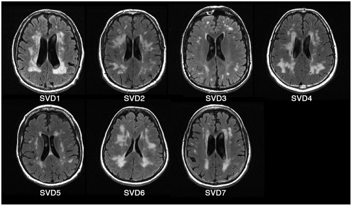
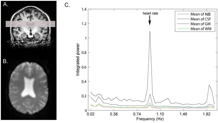
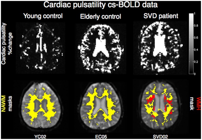
Resting state BOLD data were collected with a 250 ms repetition time (TR) to achieve unaliased, ungated cardiac sampled BOLD (cs-BOLD) images on 11 young adult controls, 10 healthy older adult controls and 7 adults with extensive white matter hyperintensities (WMH) from SVD. Tissue classes (WM and GM) were segmented on T1 images. WMH were identified on FLAIR images in the SVD group. Raw physiological noise (σphysio) and cardiac pulsatility (i.e. fluctuations at the cardiac frequency) were calculated voxel wise and group differences were tested by ANOVA. It was also possible to calculate σphysio in 2s TR cardiac aliased whole-brain BOLD (wb-BOLD) data (N = 84) obtained from the International Consortium for Brain Mapping.
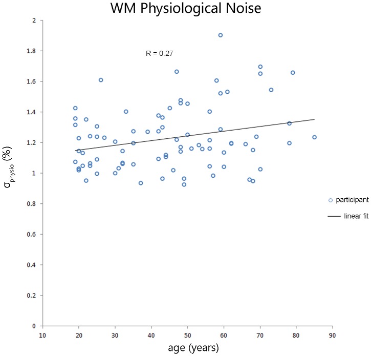
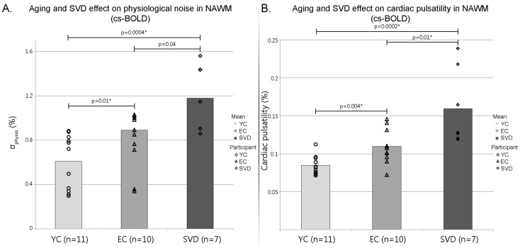
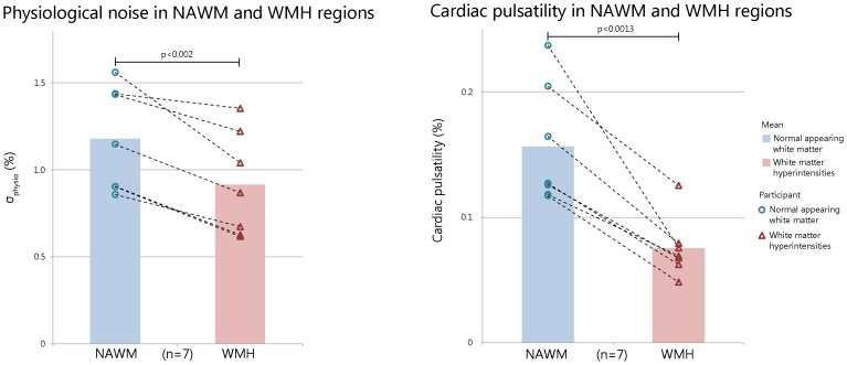
CS-BOLD metrics showed an aging and SVD effects (p<0.0005). Covariates such as thermal noise, WM volume and partial volume did not influence the significant aging effect seen on the cardiac pulsatility metric (p<0.017) but did influence the σphysio (p = 0.184). As a verification of the cs-BOLD findings, the wb-BOLD also showed a linear aging effect of σphysio in WM. In the SVD adults, cardiac pulsatility and σphysio were lower in WMH regions compared to normal appearing white matter (NAWM) regions (p<0.0013 and p<0.002, respectively). Cardiac pulsatility was better able to distinguish WMH regions from NAWM than σphysio as measured by effect size (Cohen's d 2.2 and 0.88, respectively).
NAWM was found to have graded increases in cardiac pulsations due to age and SVD, independently. Within SVD participants, WMH lesions had reduced physiological noise compared to NAWM. Cardiac pulsatility in resting BOLD data may provide a complementary dynamic measure of WM integrity to add to static FLAIR anatomical images.
确定 T2*加权血氧水平依赖(BOLD)对比图像上的白质信号波动是否与衰老和脑小血管疾病(SVD)有关。
使用 250ms 重复时间(TR)采集静息状态 BOLD 数据,以在 11 名年轻成人对照组、10 名健康老年成人对照组和 7 名 SVD 患者中获得无混淆、无门控的心脏采样 BOLD(cs-BOLD)图像。在 T1 图像上对组织分类(WM 和 GM)进行分割。在 SVD 组的 FLAIR 图像上识别出脑白质高信号(WMH)。在从国际脑映射联盟获得的 2s TR 心脏混淆全脑 BOLD(wb-BOLD)数据(N=84)中,以体素方式计算出生理噪声(σphysio)和心脏搏动(即心脏频率的波动),并通过 ANOVA 测试组间差异。也可以在 SVD 成人中,与正常表现白质(NAWM)区域相比,WMH 区域的心脏搏动和 σphysio 较低(p<0.0013 和 p<0.002,分别)。心脏搏动在区分 WMH 区域和 NAWM 方面比 σphysio 更有效,其效果大小分别为 2.2 和 0.88(Cohen's d)。
发现 NAWM 由于衰老和 SVD 而出现分级增加的心脏搏动,这是独立的。在 SVD 参与者中,与 NAWM 相比,WMH 病变的生理噪声降低。静息 BOLD 数据中的心脏搏动可能为 WM 完整性提供补充的动态测量,以补充静态 FLAIR 解剖图像。