• 文献检索
  • 文档翻译
  • 深度研究
  • 学术资讯
  • Suppr Zotero 插件Zotero 插件
  • 邀请有礼
  • 套餐&价格
  • 历史记录
应用&插件
Suppr Zotero 插件Zotero 插件浏览器插件Mac 客户端Windows 客户端微信小程序
定价
高级版会员购买积分包购买API积分包
服务
文献检索文档翻译深度研究API 文档MCP 服务
关于我们
关于 Suppr公司介绍联系我们用户协议隐私条款
关注我们

Suppr 超能文献

核心技术专利:CN118964589B侵权必究
粤ICP备2023148730 号-1Suppr @ 2026

文献检索

告别复杂PubMed语法,用中文像聊天一样搜索,搜遍4000万医学文献。AI智能推荐,让科研检索更轻松。

立即免费搜索

文件翻译

保留排版,准确专业,支持PDF/Word/PPT等文件格式,支持 12+语言互译。

免费翻译文档

深度研究

AI帮你快速写综述,25分钟生成高质量综述,智能提取关键信息,辅助科研写作。

立即免费体验

重症监护脑电监测中爆发抑制模式的实时分割。

Real-time segmentation of burst suppression patterns in critical care EEG monitoring.

机构信息

Department of Neurology, Massachusetts General Hospital, Boston, MA, USA.

出版信息

J Neurosci Methods. 2013 Sep 30;219(1):131-41. doi: 10.1016/j.jneumeth.2013.07.003. Epub 2013 Jul 23.

DOI:10.1016/j.jneumeth.2013.07.003
PMID:23891828
原文链接:https://pmc.ncbi.nlm.nih.gov/articles/PMC3939433/
Abstract

OBJECTIVE

Develop a real-time algorithm to automatically discriminate suppressions from non-suppressions (bursts) in electroencephalograms of critically ill adult patients.

METHODS

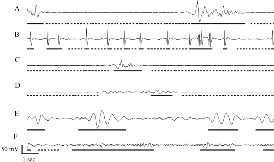
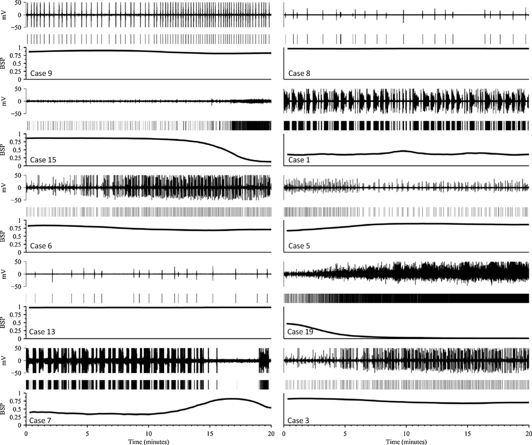
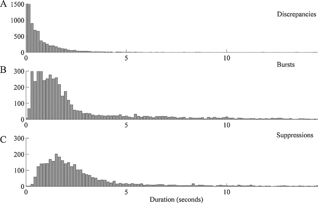
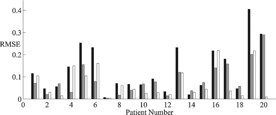
A real-time method for segmenting adult ICU EEG data into bursts and suppressions is presented based on thresholding local voltage variance. Results are validated against manual segmentations by two experienced human electroencephalographers. We compare inter-rater agreement between manual EEG segmentations by experts with inter-rater agreement between human vs automatic segmentations, and investigate the robustness of segmentation quality to variations in algorithm parameter settings. We further compare the results of using these segmentations as input for calculating the burst suppression probability (BSP), a continuous measure of depth-of-suppression.

RESULTS

Automated segmentation was comparable to manual segmentation, i.e. algorithm-vs-human agreement was comparable to human-vs-human agreement, as judged by comparing raw EEG segmentations or the derived BSP signals. Results were robust to modest variations in algorithm parameter settings.

CONCLUSIONS

Our automated method satisfactorily segments burst suppression data across a wide range adult ICU EEG patterns. Performance is comparable to or exceeds that of manual segmentation by human electroencephalographers.

SIGNIFICANCE

Automated segmentation of burst suppression EEG patterns is an essential component of quantitative brain activity monitoring in critically ill and anesthetized adults. The segmentations produced by our algorithm provide a basis for accurate tracking of suppression depth.

摘要

目的

开发一种实时算法,以自动区分危重病成人患者脑电图中的抑制(爆发)和非抑制(爆发)。

方法

提出了一种基于局部电压方差阈值的实时方法,用于将成人 ICU EEG 数据分割为爆发和抑制。结果通过两名经验丰富的人类脑电图专家的手动分割进行验证。我们比较了手动 EEG 分割的专家之间的组内一致性与人类与自动分割之间的组内一致性,并研究了分割质量对算法参数设置变化的稳健性。我们进一步比较了将这些分割作为输入用于计算爆发抑制概率(BSP)的结果,BSP 是抑制深度的连续度量。

结果

自动分割与手动分割相当,即通过比较原始 EEG 分割或衍生的 BSP 信号,算法与人类的一致性与人类与人类的一致性相当。结果对算法参数设置的适度变化具有稳健性。

结论

我们的自动方法在广泛的成人 ICU EEG 模式下令人满意地分割了爆发抑制数据。性能与人类脑电图专家的手动分割相当或超过。

意义

爆发抑制 EEG 模式的自动分割是对危重病和麻醉成人进行定量脑活动监测的重要组成部分。我们算法生成的分割为准确跟踪抑制深度提供了基础。

https://cdn.ncbi.nlm.nih.gov/pmc/blobs/c444/3939433/ef62b4fab9d8/nihms553719f5.jpg
https://cdn.ncbi.nlm.nih.gov/pmc/blobs/c444/3939433/cb5b0d2ac44e/nihms553719f1.jpg
https://cdn.ncbi.nlm.nih.gov/pmc/blobs/c444/3939433/2a14f45f169e/nihms553719f2.jpg
https://cdn.ncbi.nlm.nih.gov/pmc/blobs/c444/3939433/5fd5a5d3cdd4/nihms553719f3.jpg
https://cdn.ncbi.nlm.nih.gov/pmc/blobs/c444/3939433/76fb4999d173/nihms553719f4.jpg
https://cdn.ncbi.nlm.nih.gov/pmc/blobs/c444/3939433/ef62b4fab9d8/nihms553719f5.jpg

相似文献

1
Real-time segmentation of burst suppression patterns in critical care EEG monitoring.重症监护脑电监测中爆发抑制模式的实时分割。
J Neurosci Methods. 2013 Sep 30;219(1):131-41. doi: 10.1016/j.jneumeth.2013.07.003. Epub 2013 Jul 23.
2
Detection of EEG burst-suppression in neurocritical care patients using an unsupervised machine learning algorithm.使用无监督机器学习算法检测神经危重症患者的脑电爆发抑制。
Clin Neurophysiol. 2021 Oct;132(10):2485-2492. doi: 10.1016/j.clinph.2021.07.018. Epub 2021 Aug 5.
3
Monitoring burst suppression in critically ill patients: Multi-centric evaluation of a novel method.监测重症患者的爆发抑制:一种新方法的多中心评估
Clin Neurophysiol. 2016 Apr;127(4):2038-46. doi: 10.1016/j.clinph.2016.02.001. Epub 2016 Feb 9.
4
Automatic analysis and monitoring of burst suppression in anesthesia.麻醉中爆发抑制的自动分析与监测
J Clin Monit Comput. 2002 Feb;17(2):125-34. doi: 10.1023/a:1016393904439.
5
Automatic detection of burst suppression.爆发抑制的自动检测。
Annu Int Conf IEEE Eng Med Biol Soc. 2007;2007:553-6. doi: 10.1109/IEMBS.2007.4352350.
6
A closed-loop anesthetic delivery system for real-time control of burst suppression.实时控制爆发抑制的闭环麻醉输送系统。
J Neural Eng. 2013 Aug;10(4):046004. doi: 10.1088/1741-2560/10/4/046004. Epub 2013 Jun 7.
7
Burst suppression probability algorithms: state-space methods for tracking EEG burst suppression.突发抑制概率算法:用于跟踪脑电图突发抑制的状态空间方法。
J Neural Eng. 2013 Oct;10(5):056017. doi: 10.1088/1741-2560/10/5/056017. Epub 2013 Sep 10.
8
Burst-suppression with identical bursts: a distinct EEG pattern with poor outcome in postanoxic coma.爆发抑制伴同形态完全一致的爆发:一种与不良预后相关的独特脑电图模式,见于缺氧性昏迷后。
Clin Neurophysiol. 2014 May;125(5):947-54. doi: 10.1016/j.clinph.2013.10.017. Epub 2013 Oct 26.
9
Multi-atlas segmentation of the whole hippocampus and subfields using multiple automatically generated templates.使用多个自动生成的模板对整个海马体及其子区进行多图谱分割。
Neuroimage. 2014 Nov 1;101:494-512. doi: 10.1016/j.neuroimage.2014.04.054. Epub 2014 Apr 29.
10
Automated and manual hippocampal segmentation techniques: Comparison of results, reproducibility and clinical applicability.自动化和手动海马分割技术:结果比较、可重复性和临床适用性。
Neuroimage Clin. 2019;21:101574. doi: 10.1016/j.nicl.2018.10.012. Epub 2018 Oct 14.

引用本文的文献

1
Toward a Universal Map of EEG: A Semantic, Low-Dimensional Manifold for EEG Classification, Clustering, and Prognostication.迈向脑电图通用图谱:用于脑电图分类、聚类和预后预测的语义低维流形
Ann Neurol. 2025 Aug;98(2):357-368. doi: 10.1002/ana.27260. Epub 2025 Jun 20.
2
Spectral Power Analysis of Delta Waves in Neonatal Electroencephalography: A Tool for Assessing Brain Maturation and Injury.新生儿脑电图中δ波的频谱功率分析:一种评估脑成熟度和损伤的工具
Cureus. 2025 Mar 16;17(3):e80680. doi: 10.7759/cureus.80680. eCollection 2025 Mar.
3
Electrophysiological monitoring of neurological functions at the acute phase of brain injury: An overview of current knowledge and future perspectives in the adult population.

本文引用的文献

1
Burst suppression probability algorithms: state-space methods for tracking EEG burst suppression.突发抑制概率算法:用于跟踪脑电图突发抑制的状态空间方法。
J Neural Eng. 2013 Oct;10(5):056017. doi: 10.1088/1741-2560/10/5/056017. Epub 2013 Sep 10.
2
Continuous electroencephalography monitoring for early prediction of neurological outcome in postanoxic patients after cardiac arrest: a prospective cohort study.心脏骤停后缺氧患者的连续脑电图监测对神经功能预后的早期预测:一项前瞻性队列研究。
Crit Care Med. 2012 Oct;40(10):2867-75. doi: 10.1097/CCM.0b013e31825b94f0.
3
A neurophysiological-metabolic model for burst suppression.
脑损伤急性期神经功能的电生理监测:成年人群的当前知识概述及未来展望
Eur J Anaesthesiol Intensive Care. 2024 Feb 5;3(2):e0044. doi: 10.1097/EA9.0000000000000044. eCollection 2024 Apr.
4
Using artificial intelligence to optimize anti-seizure treatment and EEG-guided decisions in severe brain injury.利用人工智能优化重型脑损伤的抗癫痫治疗及脑电图引导决策。
Neurotherapeutics. 2025 Jan;22(1):e00524. doi: 10.1016/j.neurot.2025.e00524. Epub 2025 Jan 23.
5
The Neurostimulationist will see you now: prescribing direct electrical stimulation therapies for the human brain in epilepsy and beyond.神经刺激专家现在将为您诊治:为癫痫及其他病症的人脑开具直接电刺激疗法的处方。
Front Hum Neurosci. 2024 Sep 4;18:1439541. doi: 10.3389/fnhum.2024.1439541. eCollection 2024.
6
Do all sedatives promote biological sleep electroencephalogram patterns? A machine learning framework to identify biological sleep promoting sedatives using electroencephalogram.所有镇静剂都能促进生物睡眠脑电图模式吗?使用脑电图识别促进生物睡眠的镇静剂的机器学习框架。
PLoS One. 2024 Jul 2;19(7):e0304413. doi: 10.1371/journal.pone.0304413. eCollection 2024.
7
The Influence of Preoperative Physical Activity on Intraoperative Brain Function in Cardiac Surgical patients.术前身体活动对心脏手术患者术中脑功能的影响。
Res Sq. 2024 Jun 7:rs.3.rs-4427122. doi: 10.21203/rs.3.rs-4427122/v1.
8
Flexible, scalable, high channel count stereo-electrode for recording in the human brain.灵活、可扩展、高通道数立体电极,用于人脑记录。
Nat Commun. 2024 Jan 17;15(1):218. doi: 10.1038/s41467-023-43727-9.
9
Mapping general anesthesia states based on electro-encephalogram transition phases.基于脑电过渡相位的全身麻醉状态映射。
Neuroimage. 2024 Jan;285:120498. doi: 10.1016/j.neuroimage.2023.120498. Epub 2023 Dec 20.
10
DREDge: robust motion correction for high-density extracellular recordings across species.DREDge:跨物种高密度细胞外记录的稳健运动校正
bioRxiv. 2023 Oct 29:2023.10.24.563768. doi: 10.1101/2023.10.24.563768.
爆发抑制的神经生理代谢模型。
Proc Natl Acad Sci U S A. 2012 Feb 21;109(8):3095-100. doi: 10.1073/pnas.1121461109. Epub 2012 Feb 7.
4
A state-space model of the burst suppression ratio.爆发抑制率的状态空间模型。
Annu Int Conf IEEE Eng Med Biol Soc. 2011;2011:1431-4. doi: 10.1109/IEMBS.2011.6090354.
5
Artifact and recording concepts in EEG.脑电图中的伪迹和记录概念。
J Clin Neurophysiol. 2011 Jun;28(3):252-63. doi: 10.1097/WNP.0b013e31821c3c93.
6
General anesthesia and altered states of arousal: a systems neuroscience analysis.全身麻醉与意识改变状态:系统神经科学分析。
Annu Rev Neurosci. 2011;34:601-28. doi: 10.1146/annurev-neuro-060909-153200.
7
The treatment of status epilepticus.癫痫持续状态的治疗。
Curr Opin Neurol. 2011 Apr;24(2):165-70. doi: 10.1097/WCO.0b013e3283446f31.
8
A randomized trial for the treatment of refractory status epilepticus.一项治疗耐药性癫痫持续状态的随机试验。
Neurocrit Care. 2011 Feb;14(1):4-10. doi: 10.1007/s12028-010-9445-z.
9
Can BIS monitoring be used to assess the depth of propofol anesthesia in the treatment of refractory status epilepticus?脑电双频指数监测能否用于评估丙泊酚麻醉在治疗耐药性癫痫持续状态中的深度?
Epilepsia. 2010 Aug;51(8):1580-6. doi: 10.1111/j.1528-1167.2009.02514.x. Epub 2010 Feb 3.
10
The burst-suppression electroencephalogram.爆发抑制脑电图
Am J Electroneurodiagnostic Technol. 2009 Dec;49(4):333-41.