• 文献检索
  • 文档翻译
  • 深度研究
  • 学术资讯
  • Suppr Zotero 插件Zotero 插件
  • 邀请有礼
  • 套餐&价格
  • 历史记录
应用&插件
Suppr Zotero 插件Zotero 插件浏览器插件Mac 客户端Windows 客户端微信小程序
定价
高级版会员购买积分包购买API积分包
服务
文献检索文档翻译深度研究API 文档MCP 服务
关于我们
关于 Suppr公司介绍联系我们用户协议隐私条款
关注我们

Suppr 超能文献

核心技术专利:CN118964589B侵权必究
粤ICP备2023148730 号-1Suppr @ 2026

文献检索

告别复杂PubMed语法,用中文像聊天一样搜索,搜遍4000万医学文献。AI智能推荐,让科研检索更轻松。

立即免费搜索

文件翻译

保留排版,准确专业,支持PDF/Word/PPT等文件格式,支持 12+语言互译。

免费翻译文档

深度研究

AI帮你快速写综述,25分钟生成高质量综述,智能提取关键信息,辅助科研写作。

立即免费体验

自动化和手动海马分割技术:结果比较、可重复性和临床适用性。

Automated and manual hippocampal segmentation techniques: Comparison of results, reproducibility and clinical applicability.

机构信息

Drexel University College of Medicine, Philadelphia, PA, USA.

School of Medicine, University of California Irvine, Irvine, CA, USA.

出版信息

Neuroimage Clin. 2019;21:101574. doi: 10.1016/j.nicl.2018.10.012. Epub 2018 Oct 14.

DOI:10.1016/j.nicl.2018.10.012
PMID:30553759
原文链接:https://pmc.ncbi.nlm.nih.gov/articles/PMC6413347/
Abstract

BACKGROUND

Imaging techniques used to measure hippocampal atrophy are key to understanding the clinical progression of Alzheimer's disease (AD). Various semi-automated hippocampal segmentation techniques are available and require human expert input to learn how to accurately segment new data. Our goal was to compare 1) the performance of our automated hippocampal segmentation technique relative to manual segmentations, and 2) the performance of our automated technique when provided with a training set from two different raters. We also explored the ability of hippocampal volumes obtained using manual and automated hippocampal segmentations to predict conversion from MCI to AD.

METHODS

We analyzed 161 1.5 T T1-weighted brain magnetic resonance images (MRI) from the ADCS Donepezil/Vitamin E clinical study. All subjects carried a diagnosis of mild cognitive impairment (MCI). Three different segmentation outputs (one produced by manual tracing and two produced by a semi-automated algorithm trained with training sets developed by two raters) were compared using single measure intraclass correlation statistics (smICC). The radial distance method was used to assess each segmentation technique's ability to detect hippocampal atrophy in 3D. We then compared how well each segmentation method detected baseline hippocampal differences between MCI subjects who remained stable (MCInc) and those who converted to AD (MCIc) during the trial. Our statistical maps were corrected for multiple comparisons using permutation-based statistics with a threshold of p < .01.

RESULTS

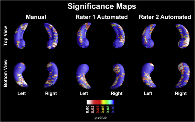
Our smICC analyses showed significant agreement between the manual and automated hippocampal segmentations from rater 1 [right smICC = 0.78 (95%CI 0.72-0.84); left smICC = 0.79 (95%CI 0.72-0.85)], the manual segmentations from rater 1 versus the automated segmentations from rater 2 [right smICC = 0.78 (95%CI 0.7-0.84); left smICC = 0.78 (95%CI 0.71-0.84)], and the automated segmentations of rater 1 versus rater 2 [right smICC = 0.97 (95%CI 0.96-0.98); left smICC = 0.97 (95%CI 0.96-0.98)]. All three segmentation methods detected significant CA1 and subicular atrophy in MCIc compared to MCInc at baseline (manual: right p = 0.0112, left p = 0.0006; automated rater 1: right p = 0.0318, left p = 0.0302; automated rater 2: right p = 0.0029, left p = 0.0166).

CONCLUSIONS

The hippocampal volumes obtained with a fast semi-automated segmentation method were highly comparable to the ones obtained with the labor-intensive manual segmentation method. The AdaBoost automated hippocampal segmentation technique is highly reliable allowing the efficient analysis of large data sets.

摘要

背景

用于测量海马体萎缩的成像技术是理解阿尔茨海默病(AD)临床进展的关键。有各种半自动海马体分割技术,需要人类专家的输入来学习如何准确地分割新数据。我们的目标是比较 1)我们的自动海马体分割技术相对于手动分割的性能,以及 2)当提供来自两个不同评分者的训练集时,我们的自动技术的性能。我们还探讨了使用手动和自动海马体分割获得的海马体体积来预测从 MCI 到 AD 的转化的能力。

方法

我们分析了来自 ADCS 多奈哌齐/维生素 E 临床研究的 161 个 1.5T T1 加权脑磁共振图像(MRI)。所有受试者均诊断为轻度认知障碍(MCI)。使用单测量组内相关统计(smICC)比较了三种不同的分割输出(一种由手动跟踪产生,两种由由两位评分者开发的训练集训练的半自动算法产生)。使用放射状距离法评估每种分割技术在 3D 中检测海马体萎缩的能力。然后,我们比较了每种分割方法在试验期间在基线时如何检测到保持稳定的 MCI 受试者(MCInc)和转化为 AD 的 MCI 受试者(MCIc)之间的海马体差异。我们的统计地图使用基于置换的统计数据进行了多重比较校正,阈值为 p<0.01。

结果

我们的 smICC 分析显示,评分者 1 的手动和自动海马体分割之间存在显著的一致性[右侧 smICC=0.78(95%CI 0.72-0.84);左侧 smICC=0.79(95%CI 0.72-0.85)],评分者 1 的手动分割与评分者 2 的自动分割之间存在显著一致性[右侧 smICC=0.78(95%CI 0.7-0.84);左侧 smICC=0.78(95%CI 0.71-0.84)],以及评分者 1 的自动分割与评分者 2 的自动分割之间存在显著一致性[右侧 smICC=0.97(95%CI 0.96-0.98);左侧 smICC=0.97(95%CI 0.96-0.98)]。与 MCInc 相比,所有三种分割方法均在基线时检测到 MCIc 的 CA1 和亚皮质萎缩(手动:右侧 p=0.0112,左侧 p=0.0006;自动评分者 1:右侧 p=0.0318,左侧 p=0.0302;自动评分者 2:右侧 p=0.0029,左侧 p=0.0166)。

结论

使用快速半自动分割方法获得的海马体体积与使用劳动密集型手动分割方法获得的体积高度可比。AdaBoost 自动海马体分割技术非常可靠,允许对大型数据集进行高效分析。

https://cdn.ncbi.nlm.nih.gov/pmc/blobs/2b1f/6413347/90ffd0b47279/gr1.jpg

相似文献

1
Automated and manual hippocampal segmentation techniques: Comparison of results, reproducibility and clinical applicability.自动化和手动海马分割技术:结果比较、可重复性和临床适用性。
Neuroimage Clin. 2019;21:101574. doi: 10.1016/j.nicl.2018.10.012. Epub 2018 Oct 14.
2
Hippocampal volume change measurement: quantitative assessment of the reproducibility of expert manual outlining and the automated methods FreeSurfer and FIRST.海马体积变化测量:专家手动勾勒以及自动化方法FreeSurfer和FIRST的可重复性定量评估。
Neuroimage. 2014 May 15;92:169-81. doi: 10.1016/j.neuroimage.2014.01.058. Epub 2014 Feb 9.
3
Training labels for hippocampal segmentation based on the EADC-ADNI harmonized hippocampal protocol.基于 EADC-ADNI 协调的海马协议的海马分割训练标签。
Alzheimers Dement. 2015 Feb;11(2):175-83. doi: 10.1016/j.jalz.2014.12.002. Epub 2015 Jan 20.
4
Automated 3D mapping of hippocampal atrophy and its clinical correlates in 400 subjects with Alzheimer's disease, mild cognitive impairment, and elderly controls.对400名患有阿尔茨海默病、轻度认知障碍的受试者以及老年对照者进行海马萎缩的自动三维测绘及其临床相关性研究。
Hum Brain Mapp. 2009 Sep;30(9):2766-88. doi: 10.1002/hbm.20708.
5
In vivo analysis of hippocampal subfield atrophy in mild cognitive impairment via semi-automatic segmentation of T2-weighted MRI.利用 T2 加权 MRI 半自动分割分析轻度认知障碍患者海马亚区萎缩
J Alzheimers Dis. 2012;31(1):85-99. doi: 10.3233/JAD-2012-111931.
6
Multi-atlas segmentation of the whole hippocampus and subfields using multiple automatically generated templates.使用多个自动生成的模板对整个海马体及其子区进行多图谱分割。
Neuroimage. 2014 Nov 1;101:494-512. doi: 10.1016/j.neuroimage.2014.04.054. Epub 2014 Apr 29.
7
Structural magnetic resonance imaging for the early diagnosis of dementia due to Alzheimer's disease in people with mild cognitive impairment.用于轻度认知障碍患者阿尔茨海默病所致痴呆早期诊断的结构磁共振成像
Cochrane Database Syst Rev. 2020 Mar 2;3(3):CD009628. doi: 10.1002/14651858.CD009628.pub2.
8
Conversion of mild cognitive impairment to Alzheimer disease predicted by hippocampal atrophy maps.海马萎缩图谱预测轻度认知障碍向阿尔茨海默病的转化
Arch Neurol. 2006 May;63(5):693-9. doi: 10.1001/archneur.63.5.693.
9
Non-linear registration improves statistical power to detect hippocampal atrophy in aging and dementia.非线性配准提高了检测衰老和痴呆中海马体萎缩的统计功效。
Neuroimage Clin. 2019;23:101902. doi: 10.1016/j.nicl.2019.101902. Epub 2019 Jun 18.
10
Semi-automated hippocampal segmentation in people with cognitive impairment using an age appropriate template for registration.使用适合年龄的配准模板对认知障碍患者进行半自动海马体分割。
J Magn Reson Imaging. 2015 Dec;42(6):1631-8. doi: 10.1002/jmri.24966. Epub 2015 Jul 3.

引用本文的文献

1
Hippocampal subfield volumes in mild cognitive impairment and alzheimer's disease: a systematic review and meta-analysis.轻度认知障碍和阿尔茨海默病患者的海马亚区体积:系统评价和荟萃分析。
Brain Imaging Behav. 2023 Dec;17(6):778-793. doi: 10.1007/s11682-023-00804-3. Epub 2023 Sep 28.
2
Automated identification of piglet brain tissue from MRI images using Region-based Convolutional Neural Networks.基于区域卷积神经网络的 MRI 图像中小猪脑组织的自动识别。
PLoS One. 2023 May 11;18(5):e0284951. doi: 10.1371/journal.pone.0284951. eCollection 2023.
3
Deep Learning Based Detection of Enlarged Perivascular Spaces on Brain MRI.

本文引用的文献

1
Defining SNAP by cross-sectional and longitudinal definitions of neurodegeneration.根据神经退行性变的横断面和纵向定义来定义 SNAP。
Neuroimage Clin. 2018 Feb 8;18:407-412. doi: 10.1016/j.nicl.2018.02.008. eCollection 2018.
2
Mild Cognitive Impairment.轻度认知障碍
Continuum (Minneap Minn). 2016 Apr;22(2 Dementia):404-18. doi: 10.1212/CON.0000000000000313.
3
Comparing 3T and 1.5T MRI for mapping hippocampal atrophy in the Alzheimer's Disease Neuroimaging Initiative.在阿尔茨海默病神经影像倡议中比较3T和1.5T磁共振成像用于绘制海马萎缩情况
基于深度学习的脑磁共振成像上血管周围间隙增宽的检测
Neuroimage Rep. 2023 Mar;3(1). doi: 10.1016/j.ynirp.2023.100162. Epub 2023 Mar 7.
4
Deep Learning Segmentation of Triple-Negative Breast Cancer (TNBC) Patient Derived Tumor Xenograft (PDX) and Sensitivity of Radiomic Pipeline to Tumor Probability Boundary.三阴性乳腺癌(TNBC)患者来源肿瘤异种移植(PDX)的深度学习分割及影像组学流程对肿瘤概率边界的敏感性
Cancers (Basel). 2021 Jul 28;13(15):3795. doi: 10.3390/cancers13153795.
5
Diagnostic Performance of Automated MRI Volumetry by icobrain dm for Alzheimer's Disease in a Clinical Setting: A REMEMBER Study.icobrain dm 自动化 MRI 容积测量技术在临床环境下对阿尔茨海默病的诊断性能:REMEMBER 研究。
J Alzheimers Dis. 2021;83(2):623-639. doi: 10.3233/JAD-210450.
6
Imaging Techniques in Alzheimer's Disease: A Review of Applications in Early Diagnosis and Longitudinal Monitoring.阿尔茨海默病的影像学技术:在早期诊断和纵向监测中的应用综述。
Int J Mol Sci. 2021 Feb 20;22(4):2110. doi: 10.3390/ijms22042110.
7
Inflammation, hippocampal volume, and cognition in schizophrenia: results from the Northern Finland Birth Cohort 1966.精神分裂症中的炎症、海马体体积和认知:来自 1966 年芬兰北部出生队列的研究结果。
Eur Arch Psychiatry Clin Neurosci. 2021 Jun;271(4):609-622. doi: 10.1007/s00406-020-01134-x. Epub 2020 May 7.
8
Imaging biomarkers in neurodegeneration: current and future practices.神经退行性疾病的影像学生物标志物:当前和未来的实践。
Alzheimers Res Ther. 2020 Apr 27;12(1):49. doi: 10.1186/s13195-020-00612-7.
AJNR Am J Neuroradiol. 2015 Apr;36(4):653-60. doi: 10.3174/ajnr.A4228. Epub 2015 Jan 22.
4
Harmonized benchmark labels of the hippocampus on magnetic resonance: the EADC-ADNI project.磁共振海马的协调基准标签:EADC-ADNI 项目。
Alzheimers Dement. 2015 Feb;11(2):151-60.e5. doi: 10.1016/j.jalz.2013.12.019. Epub 2014 Sep 13.
5
Automated voxel-by-voxel tissue classification for hippocampal segmentation: methods and validation.用于海马体分割的逐体素自动组织分类:方法与验证
Phys Med. 2014 Dec;30(8):878-87. doi: 10.1016/j.ejmp.2014.06.044. Epub 2014 Jul 11.
6
Comparing new templates and atlas-based segmentations in the volumetric analysis of brain magnetic resonance images for diagnosing Alzheimer's disease.比较新模板和基于图谱的脑磁共振图像容积分析在阿尔茨海默病诊断中的分割。
Alzheimers Dement. 2012 Sep;8(5):399-406. doi: 10.1016/j.jalz.2011.07.002.
7
Hippocampal atrophy and ventricular enlargement in normal aging, mild cognitive impairment (MCI), and Alzheimer Disease.海马萎缩和脑室扩大在正常衰老、轻度认知障碍(MCI)和阿尔茨海默病中。
Alzheimer Dis Assoc Disord. 2012 Jan-Mar;26(1):17-27. doi: 10.1097/WAD.0b013e3182163b62.
8
Appearance-based modeling for segmentation of hippocampus and amygdala using multi-contrast MR imaging.基于外观的多对比度磁共振成像分割海马体和杏仁核的方法。
Neuroimage. 2011 Sep 15;58(2):549-59. doi: 10.1016/j.neuroimage.2011.06.054. Epub 2011 Jun 25.
9
Automatic morphometry in Alzheimer's disease and mild cognitive impairment.阿尔茨海默病和轻度认知障碍的自动形态计量学。
Neuroimage. 2011 Jun 15;56(4):2024-37. doi: 10.1016/j.neuroimage.2011.03.014. Epub 2011 Mar 11.
10
Dataset of magnetic resonance images of nonepileptic subjects and temporal lobe epilepsy patients for validation of hippocampal segmentation techniques.无癫痫发作受试者和颞叶癫痫患者的磁共振图像数据集,用于验证海马分割技术。
Neuroinformatics. 2011 Dec;9(4):335-46. doi: 10.1007/s12021-010-9096-4.