• 文献检索
  • 文档翻译
  • 深度研究
  • 学术资讯
  • Suppr Zotero 插件Zotero 插件
  • 邀请有礼
  • 套餐&价格
  • 历史记录
应用&插件
Suppr Zotero 插件Zotero 插件浏览器插件Mac 客户端Windows 客户端微信小程序
定价
高级版会员购买积分包购买API积分包
服务
文献检索文档翻译深度研究API 文档MCP 服务
关于我们
关于 Suppr公司介绍联系我们用户协议隐私条款
关注我们

Suppr 超能文献

核心技术专利:CN118964589B侵权必究
粤ICP备2023148730 号-1Suppr @ 2026

文献检索

告别复杂PubMed语法,用中文像聊天一样搜索,搜遍4000万医学文献。AI智能推荐,让科研检索更轻松。

立即免费搜索

文件翻译

保留排版,准确专业,支持PDF/Word/PPT等文件格式,支持 12+语言互译。

免费翻译文档

深度研究

AI帮你快速写综述,25分钟生成高质量综述,智能提取关键信息,辅助科研写作。

立即免费体验

相似文献

1
Fe5C2 nanoparticles with high MRI contrast enhancement for tumor imaging.具有高磁共振成像对比增强效果的Fe5C2纳米颗粒用于肿瘤成像。
Small. 2014 Apr 9;10(7):1245-9. doi: 10.1002/smll.201303263. Epub 2013 Dec 18.
2
Casein-Coated Fe5C2 Nanoparticles with Superior r2 Relaxivity for Liver-Specific Magnetic Resonance Imaging.具有优异 r2 弛豫率的酪蛋白包覆 Fe5C2 纳米颗粒用于肝脏特异性磁共振成像。
Theranostics. 2015 Aug 9;5(11):1225-32. doi: 10.7150/thno.12570. eCollection 2015.
3
Highly magnetic iron carbide nanoparticles as effective T(2) contrast agents.高磁性碳化铁纳米颗粒作为有效的 T(2)对比剂。
Nanoscale. 2014 Jan 21;6(2):726-30. doi: 10.1039/c3nr04691e.
4
Magnetic Reactive Oxygen Species Nanoreactor for Switchable Magnetic Resonance Imaging Guided Cancer Therapy Based on pH-Sensitive FeC@FeO Nanoparticles.基于 pH 敏感的 FeC@FeO 纳米粒子的可切换磁共振成像引导癌症治疗的磁性反应氧物种纳米反应器。
ACS Nano. 2019 Sep 24;13(9):10002-10014. doi: 10.1021/acsnano.9b01740. Epub 2019 Aug 26.
5
Monodisperse Au-FeC Janus Nanoparticles: An Attractive Multifunctional Material for Triple-Modal Imaging-Guided Tumor Photothermal Therapy.单分散 Au-FeC 手性纳米粒子:一种用于三模态成像引导肿瘤光热治疗的有吸引力的多功能材料。
ACS Nano. 2017 Sep 26;11(9):9239-9248. doi: 10.1021/acsnano.7b04461. Epub 2017 Sep 5.
6
Magnetic Targeting, Tumor Microenvironment-Responsive Intelligent Nanocatalysts for Enhanced Tumor Ablation.磁性靶向、肿瘤微环境响应智能纳米催化剂增强肿瘤消融。
ACS Nano. 2018 Nov 27;12(11):11000-11012. doi: 10.1021/acsnano.8b05042. Epub 2018 Oct 25.
7
Increased transverse relaxivity in ultrasmall superparamagnetic iron oxide nanoparticles used as MRI contrast agent for biomedical imaging.用作生物医学成像磁共振成像造影剂的超小超顺磁性氧化铁纳米颗粒中横向弛豫率增加。
Contrast Media Mol Imaging. 2016 Sep;11(5):350-361. doi: 10.1002/cmmi.1698. Epub 2016 May 27.
8
Biocompatible Peptide-Coated Ultrasmall Superparamagnetic Iron Oxide Nanoparticles for In Vivo Contrast-Enhanced Magnetic Resonance Imaging.用于体内对比增强磁共振成像的生物相容型肽涂层超顺磁性氧化铁纳米颗粒。
ACS Nano. 2018 Jul 24;12(7):6480-6491. doi: 10.1021/acsnano.7b07572. Epub 2018 Jul 11.
9
Comparative analysis of the 1H NMR relaxation enhancement produced by iron oxide and core-shell iron-iron oxide nanoparticles.氧化铁和核壳型铁-氧化铁纳米颗粒产生的1H NMR弛豫增强的比较分析。
Magn Reson Imaging. 2007 Dec;25(10):1437-41. doi: 10.1016/j.mri.2007.04.006. Epub 2007 Jun 13.
10
Upconverting rare-earth nanoparticles with a paramagnetic lanthanide complex shell for upconversion fluorescent and magnetic resonance dual-modality imaging.上转换稀土纳米粒子与顺磁镧系元素配合物壳层用于上转换荧光和磁共振双模式成像。
Nanotechnology. 2013 May 3;24(17):175101. doi: 10.1088/0957-4484/24/17/175101. Epub 2013 Apr 4.

引用本文的文献

1
Principles and applications of magnetic nanomaterials in magnetically guided bioimaging.磁性纳米材料在磁导向生物成像中的原理与应用
Mater Today Phys. 2023 Mar;32. doi: 10.1016/j.mtphys.2023.101003. Epub 2023 Feb 2.
2
One-Step Low-Temperature Synthesis of Metastable ε-Iron Carbide Nanoparticles with Unique Catalytic Properties Beyond Conventional Iron Catalysts.具有超越传统铁催化剂独特催化性能的亚稳ε-碳化铁纳米颗粒的一步低温合成。
Small. 2025 Jul;21(27):e2412217. doi: 10.1002/smll.202412217. Epub 2025 Apr 24.
3
Surface Engineering of Magnetic Iron Oxide Nanoparticles for Breast Cancer Diagnostics and Drug Delivery.磁性氧化铁纳米粒子的表面工程用于乳腺癌诊断和药物输送。
Int J Nanomedicine. 2024 Aug 17;19:8437-8461. doi: 10.2147/IJN.S477652. eCollection 2024.
4
In Vitro Toxicological Insights from the Biomedical Applications of Iron Carbide Nanoparticles in Tumor Theranostics: A Systematic Review and Meta-Analysis.碳化铁纳米颗粒在肿瘤诊疗中的生物医学应用的体外毒理学见解:一项系统综述和荟萃分析
Nanomaterials (Basel). 2024 Apr 23;14(9):734. doi: 10.3390/nano14090734.
5
New Types of Magnetic Nanoparticles for Stimuli-Responsive Theranostic Nanoplatforms.新型磁纳米粒子用于刺激响应型治疗诊断纳米平台。
Adv Sci (Weinh). 2024 Feb;11(8):e2305459. doi: 10.1002/advs.202305459. Epub 2023 Nov 21.
6
An ultra-sensitive -weighted MR contrast agent based on Gd ion chelated FeO nanoparticles.一种基于钆离子螯合的FeO纳米颗粒的超灵敏加权磁共振造影剂。
RSC Adv. 2020 May 12;10(31):18054-18061. doi: 10.1039/d0ra01807d. eCollection 2020 May 10.
7
Environmental STEM Study of the Oxidation Mechanism for Iron and Iron Carbide Nanoparticles.铁和碳化铁纳米颗粒氧化机制的环境STEM研究
Materials (Basel). 2022 Feb 18;15(4):1557. doi: 10.3390/ma15041557.
8
Red Blood Cell-Facilitated Photodynamic Therapy for Cancer Treatment.红细胞辅助光动力疗法用于癌症治疗
Adv Funct Mater. 2016 Mar 15;26(11):1757-1768. doi: 10.1002/adfm.201504803. Epub 2016 Feb 3.
9
Water-Dispersible and Biocompatible Iron Carbide Nanoparticles with High Specific Absorption Rate.水散性和生物相容性的高比吸收率碳化铁纳米颗粒。
ACS Nano. 2019 Mar 26;13(3):2870-2878. doi: 10.1021/acsnano.8b05671. Epub 2019 Mar 7.
10
Iron Carbide@Carbon Nanocomposites: A Tool Box of Functional Materials.碳化铁@碳纳米复合材料:功能材料的工具箱
Materials (Basel). 2019 Jan 21;12(2):323. doi: 10.3390/ma12020323.

本文引用的文献

1
Octapod iron oxide nanoparticles as high-performance T₂ contrast agents for magnetic resonance imaging.八足氧化铁纳米颗粒作为高性能 T₂ 磁共振成像对比剂。
Nat Commun. 2013;4:2266. doi: 10.1038/ncomms3266.
2
Fe5C2 nanoparticles: a facile bromide-induced synthesis and as an active phase for Fischer-Tropsch synthesis.Fe5C2 纳米颗粒:一种简便的溴化物诱导合成方法及其在费托合成中的活性相。
J Am Chem Soc. 2012 Sep 26;134(38):15814-21. doi: 10.1021/ja305048p. Epub 2012 Sep 12.
3
Improving the magnetic resonance imaging contrast and detection methods with engineered magnetic nanoparticles.利用工程化磁性纳米颗粒提高磁共振成像对比度和检测方法。
Theranostics. 2012;2(1):86-102. doi: 10.7150/thno.4006. Epub 2012 Jan 10.
4
Targeting strategies for multifunctional nanoparticles in cancer imaging and therapy.多功能纳米颗粒在癌症成像与治疗中的靶向策略
Theranostics. 2012;2(1):3-44. doi: 10.7150/thno.3463. Epub 2012 Jan 1.
5
Predictive toxicology of cobalt nanoparticles and ions: comparative in vitro study of different cellular models using methods of knowledge discovery from data.钴纳米粒子和离子的预测毒理学:使用数据知识发现方法对不同细胞模型的体外比较研究。
Toxicol Sci. 2011 Aug;122(2):489-501. doi: 10.1093/toxsci/kfr124. Epub 2011 May 20.
6
Surface-engineered magnetic nanoparticle platforms for cancer imaging and therapy.表面工程磁性纳米颗粒平台用于癌症成像和治疗。
Acc Chem Res. 2011 Oct 18;44(10):883-92. doi: 10.1021/ar200044b. Epub 2011 May 6.
7
Multimodality Imaging of Integrin α(v)β(3) Expression.整合素 α(v)β(3)表达的多模态成像。
Theranostics. 2011 Feb 10;1:135-48. doi: 10.7150/thno/v01p0135.
8
Fine tuning of the relaxometry of γ-Fe2O3@SiO2 nanoparticles by tweaking the silica coating thickness.通过调整二氧化硅涂层厚度来优化 γ-Fe2O3@SiO2 纳米颗粒的弛豫率。
ACS Nano. 2010 Sep 28;4(9):5339-49. doi: 10.1021/nn101129r.
9
Nanoparticle-based theranostic agents.基于纳米粒子的治疗诊断一体化试剂。
Adv Drug Deliv Rev. 2010 Aug 30;62(11):1064-79. doi: 10.1016/j.addr.2010.07.009. Epub 2010 Aug 4.
10
Iron carbide: an ancient advanced material.碳化铁:一种古老的先进材料。
Small. 2010 Sep 6;6(17):1859-62. doi: 10.1002/smll.201000437.

具有高磁共振成像对比增强效果的Fe5C2纳米颗粒用于肿瘤成像。

Fe5C2 nanoparticles with high MRI contrast enhancement for tumor imaging.

作者信息

Tang Wei, Zhen Zipeng, Yang Ce, Wang Luning, Cowger Taku, Chen Hongmin, Todd Trever, Hekmatyar Khan, Zhao Qun, Hou Yanglong, Xie Jin

机构信息

Department of Chemistry and Bio-Imaging, Research Center (BIRC), University of Georgia, Athens, GA, 30602, USA.

出版信息

Small. 2014 Apr 9;10(7):1245-9. doi: 10.1002/smll.201303263. Epub 2013 Dec 18.

DOI:10.1002/smll.201303263
PMID:24352976
原文链接:https://pmc.ncbi.nlm.nih.gov/articles/PMC6398947/
Abstract

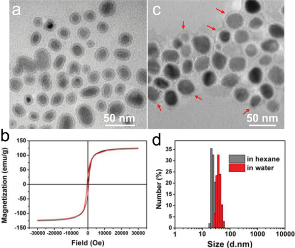
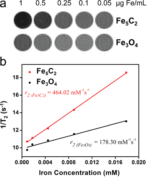
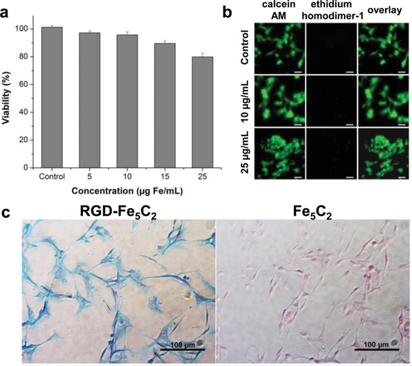
An ancient material for magnetic resonance (MR) imaging: For the first time, Fe5C2 is prepared as colloidal stable nanoparticles with good aqueous stability. The nanoparticles boast strong magnetization, excellent chemical inertness, low toxicity, and one of the highest r2 relaxivities reported to date. These nanoparticles hold great potential in MR imaging as well as in other biomedical areas.

摘要

一种用于磁共振成像的古老材料

首次将Fe5C2制备成具有良好水稳定性的胶体稳定纳米颗粒。这些纳米颗粒具有强磁化性、优异的化学惰性、低毒性以及迄今为止报道的最高r2弛豫率之一。这些纳米颗粒在磁共振成像以及其他生物医学领域具有巨大潜力。

https://cdn.ncbi.nlm.nih.gov/pmc/blobs/14d4/6398947/35279595df5c/nihms-1001407-f0004.jpg
https://cdn.ncbi.nlm.nih.gov/pmc/blobs/14d4/6398947/8b08081ff40e/nihms-1001407-f0001.jpg
https://cdn.ncbi.nlm.nih.gov/pmc/blobs/14d4/6398947/fc50aa8a3147/nihms-1001407-f0002.jpg
https://cdn.ncbi.nlm.nih.gov/pmc/blobs/14d4/6398947/22deba510abf/nihms-1001407-f0003.jpg
https://cdn.ncbi.nlm.nih.gov/pmc/blobs/14d4/6398947/35279595df5c/nihms-1001407-f0004.jpg