• 文献检索
  • 文档翻译
  • 深度研究
  • 学术资讯
  • Suppr Zotero 插件Zotero 插件
  • 邀请有礼
  • 套餐&价格
  • 历史记录
应用&插件
Suppr Zotero 插件Zotero 插件浏览器插件Mac 客户端Windows 客户端微信小程序
定价
高级版会员购买积分包购买API积分包
服务
文献检索文档翻译深度研究API 文档MCP 服务
关于我们
关于 Suppr公司介绍联系我们用户协议隐私条款
关注我们

Suppr 超能文献

核心技术专利:CN118964589B侵权必究
粤ICP备2023148730 号-1Suppr @ 2026

文献检索

告别复杂PubMed语法,用中文像聊天一样搜索,搜遍4000万医学文献。AI智能推荐,让科研检索更轻松。

立即免费搜索

文件翻译

保留排版,准确专业,支持PDF/Word/PPT等文件格式,支持 12+语言互译。

免费翻译文档

深度研究

AI帮你快速写综述,25分钟生成高质量综述,智能提取关键信息,辅助科研写作。

立即免费体验

卵巢癌细胞的侵袭性与 Let-7 miRNA 和 miR-200 外泌体分离的不协调性有关。

Ovarian cancer cell invasiveness is associated with discordant exosomal sequestration of Let-7 miRNA and miR-200.

机构信息

Centre for Clinical Diagnostics, Royal Brisbane and Women's Hospital, University of Queensland Centre for Clinical Research, Building 71/918, Herston, Queensland 4029, Australia.

出版信息

J Transl Med. 2014 Jan 6;12:4. doi: 10.1186/1479-5876-12-4.

DOI:10.1186/1479-5876-12-4
PMID:24393345
原文链接:https://pmc.ncbi.nlm.nih.gov/articles/PMC3896684/
Abstract

BACKGROUND

The role of exosomes in the pathogenesis and metastatic spread of cancer remains to be fully elucidated. Recent studies support the hypothesis that the release of exosomes from cells modifies local extracellular conditions to promote cell growth and neovascularisation. In addition, exosomes may modify the phenotype of parent and/or target cell. For example, sequestration of signaling mediators into exosomes may reduce their intracellular bioavailability to the parent cell thereby altering cell phenotype and metastatic potential. The fusion of released exosomes with target cell and delivery may also modify cell function and activity. In this study, to further elucidate the role of exosomes in ovarian cancer, the release of exosomes from two ovarian cancer cell lines of different invasive capacity and their miRNA content of exosomes were compared. The hypothesis to be tested was that ovarian cancer cell invasiveness is associated with altered release of exosomes and discordant exosomal sequestration of miRNA.

METHODS

High (SKOV-3) and low (OVCAR-3) invasive ovarian cancer cell lines were used to characterize their exosome release. SKOV-3 and OVCAR-3 cells were cultured (DMEM, 20% exosome-free FBS) under an atmosphere of 8% O2 for 24 hours. Cell-conditioned media were collected and exosomes were isolated by differential and buoyant density centrifugation and characterised by Western blot (CD63 and CD9). Exosomal microRNA (let-7a-f and miR-200a-c) content was established by real-time PCR.

RESULTS

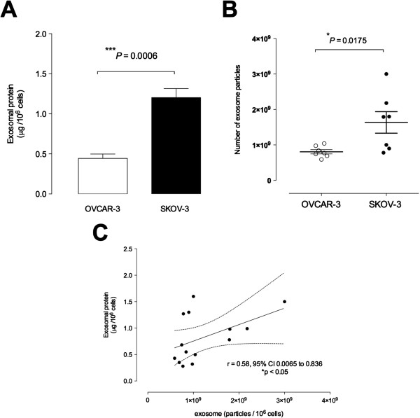
Exosomes were identified with by the presence of typical cup-shaped spherical vesicle and the expression of exosome markers: CD63, CD9. SKOV-3 cells released 2.7-fold more exosomes (1.22 ± 0.11 μg/106 cells) compared to OVCAR-3 (0.44 ± 0.05 μg/106 cells). The let-7 family miRNA transcripts were identified in both ovarian cancer cell lines and their exosomes. The let-7 family transcripts were more abundant in OVCAR-3 cell than SKOV-3 cells. In contrast, let-7 family transcripts were more abundant in exosomes from SKOV-3 than OVCAR-3. miR-200 family transcripts were only identified in OVCAR-3 cells and their exosomes.

CONCLUSIONS

The data obtained in this study are consistent with the hypothesis that the releases of exosomes varies significantly between ovarian cancer cell lines and correlates with their invasive potential.

摘要

背景

外泌体在癌症的发病机制和转移扩散中的作用仍有待充分阐明。最近的研究支持这样一种假设,即细胞释放的外泌体改变局部细胞外环境,促进细胞生长和新生血管形成。此外,外泌体还可以改变亲本和/或靶细胞的表型。例如,将信号介质隔离到外泌体中可能会降低其在亲本细胞中的细胞内生物利用度,从而改变细胞表型和转移潜能。释放的外泌体与靶细胞融合和传递也可能改变细胞功能和活性。在这项研究中,为了进一步阐明外泌体在卵巢癌中的作用,我们比较了两种侵袭能力不同的卵巢癌细胞系释放的外泌体及其外泌体中的 miRNA 含量。要检验的假设是卵巢癌细胞的侵袭能力与外泌体释放的改变以及 miRNA 的外泌体隔离不一致有关。

方法

使用高(SKOV-3)和低(OVCAR-3)侵袭性卵巢癌细胞系来表征它们的外泌体释放。将 SKOV-3 和 OVCAR-3 细胞在 8%氧气的环境下用含有 20%无外泌体胎牛血清的 DMEM 培养基培养 24 小时。收集细胞条件培养基,并通过差速和浮力密度离心分离外泌体,并通过 Western blot(CD63 和 CD9)进行鉴定。通过实时 PCR 确定外泌体 microRNA(let-7a-f 和 miR-200a-c)含量。

结果

通过存在典型的杯形球形囊泡和外泌体标记物(CD63、CD9)鉴定出外泌体。与 OVCAR-3(0.44±0.05μg/106 细胞)相比,SKOV-3 细胞释放的外泌体多 2.7 倍(1.22±0.11μg/106 细胞)。卵巢癌细胞系及其外泌体中均鉴定出 let-7 家族 miRNA 转录物。与 SKOV-3 细胞相比,OVCAR-3 细胞中的 let-7 家族转录物更为丰富。相比之下,SKOV-3 细胞中外泌体中的 let-7 家族转录物更为丰富。miR-200 家族转录物仅在 OVCAR-3 细胞及其外泌体中被鉴定。

结论

本研究获得的数据与这样一种假设一致,即外泌体的释放在卵巢癌细胞系之间差异显著,与它们的侵袭能力相关。

https://cdn.ncbi.nlm.nih.gov/pmc/blobs/6a98/3896684/e68cdd29fcec/1479-5876-12-4-6.jpg
https://cdn.ncbi.nlm.nih.gov/pmc/blobs/6a98/3896684/f76c91e7cd96/1479-5876-12-4-1.jpg
https://cdn.ncbi.nlm.nih.gov/pmc/blobs/6a98/3896684/60bd5c3a6023/1479-5876-12-4-2.jpg
https://cdn.ncbi.nlm.nih.gov/pmc/blobs/6a98/3896684/bb11b35d2a58/1479-5876-12-4-3.jpg
https://cdn.ncbi.nlm.nih.gov/pmc/blobs/6a98/3896684/a281a9c908ae/1479-5876-12-4-4.jpg
https://cdn.ncbi.nlm.nih.gov/pmc/blobs/6a98/3896684/2920a94de03d/1479-5876-12-4-5.jpg
https://cdn.ncbi.nlm.nih.gov/pmc/blobs/6a98/3896684/e68cdd29fcec/1479-5876-12-4-6.jpg

相似文献

1
Ovarian cancer cell invasiveness is associated with discordant exosomal sequestration of Let-7 miRNA and miR-200.卵巢癌细胞的侵袭性与 Let-7 miRNA 和 miR-200 外泌体分离的不协调性有关。
J Transl Med. 2014 Jan 6;12:4. doi: 10.1186/1479-5876-12-4.
2
Exosomal miR-99a-5p is elevated in sera of ovarian cancer patients and promotes cancer cell invasion by increasing fibronectin and vitronectin expression in neighboring peritoneal mesothelial cells.外泌体 miR-99a-5p 在卵巢癌患者血清中升高,并通过增加邻近腹膜间皮细胞中纤连蛋白和 vitronectin 的表达促进癌细胞侵袭。
BMC Cancer. 2018 Nov 5;18(1):1065. doi: 10.1186/s12885-018-4974-5.
3
Adipocyte derived exosomes promote cell invasion and challenge paclitaxel efficacy in ovarian cancer.脂肪细胞衍生的外泌体促进卵巢癌细胞侵袭并挑战紫杉醇疗效。
Cell Commun Signal. 2024 Sep 16;22(1):443. doi: 10.1186/s12964-024-01806-4.
4
[Screening and identification of potential miRNA involved in ovarian cancer invasion and metastasis].[参与卵巢癌侵袭和转移的潜在微小RNA的筛选与鉴定]
Zhonghua Zhong Liu Za Zhi. 2010 Sep;32(9):650-4.
5
Proteomic analysis of exosomes reveals an association between cell invasiveness and exosomal bioactivity on endothelial and mesenchymal cell migration .外泌体的蛋白质组学分析揭示了细胞侵袭性与外泌体生物活性在血管内皮细胞和间充质细胞迁移中的关联。
Clin Sci (Lond). 2018 Sep 18;132(18):2029-2044. doi: 10.1042/CS20180425. Print 2018 Sep 28.
6
Ovarian cancer-derived exosomes promote tumour metastasis : an effect modulated by the invasiveness capacity of their originating cells.卵巢癌细胞衍生的外泌体促进肿瘤转移:这种作用可被其起源细胞的侵袭能力所调节。
Clin Sci (Lond). 2019 Jul 5;133(13):1401-1419. doi: 10.1042/CS20190082. Print 2019 Jul 15.
7
TWEAK-stimulated macrophages inhibit metastasis of epithelial ovarian cancer via exosomal shuttling of microRNA.肿瘤坏死因子样弱凋亡诱导因子(TWEAK)刺激的巨噬细胞通过外泌体介导的微小RNA转运抑制上皮性卵巢癌转移。
Cancer Lett. 2017 May 1;393:60-67. doi: 10.1016/j.canlet.2017.02.009. Epub 2017 Feb 16.
8
Macrophages derived exosomes deliver miR-223 to epithelial ovarian cancer cells to elicit a chemoresistant phenotype.巨噬细胞衍生的外泌体将 miR-223 递送至上皮性卵巢癌细胞以引发耐药表型。
J Exp Clin Cancer Res. 2019 Feb 15;38(1):81. doi: 10.1186/s13046-019-1095-1.
9
Exosomal miRNA confers chemo resistance via targeting Cav1/p-gp/M2-type macrophage axis in ovarian cancer.外泌体 miRNA 通过靶向 Cav1/p-gp/M2 型巨噬细胞轴赋予卵巢癌细胞化疗耐药性。
EBioMedicine. 2018 Dec;38:100-112. doi: 10.1016/j.ebiom.2018.11.004. Epub 2018 Nov 25.
10
Protocol for serum exosomal miRNAs analysis in prostate cancer patients treated with radiotherapy.前列腺癌患者放射治疗后血清外泌体 miRNA 分析方案。
J Transl Med. 2018 Aug 13;16(1):223. doi: 10.1186/s12967-018-1592-6.

引用本文的文献

1
Long Non-Coding, Micro, and Circular RNAs in Ovarian Cancer Metastasis: Pathways and Treatment Approaches.长链非编码RNA、微小RNA和环状RNA在卵巢癌转移中的作用:途径与治疗方法
Reprod Sci. 2025 Aug 15. doi: 10.1007/s43032-025-01948-x.
2
Investigating the Effects of ONC206 Alone and in Combination with Cisplatin on Ovarian Cancer Cell Models.研究ONC206单独及与顺铂联合对卵巢癌细胞模型的影响。
Curr Issues Mol Biol. 2025 Jun 12;47(6):451. doi: 10.3390/cimb47060451.
3
Extracellular vesicle-associated miR-515-5p from adipose tissue regulates placental metabolism and fetal growth in gestational diabetes mellitus.

本文引用的文献

1
Exosomes in Plasma of Patients with Ovarian Carcinoma: Potential Biomarkers of Tumor Progression and Response to Therapy.卵巢癌患者血浆中的外泌体:肿瘤进展和治疗反应的潜在生物标志物
Gynecol Obstet (Sunnyvale). 2013 Apr 29;Suppl 4:3. doi: 10.4172/2161-0932.S4-003.
2
Hypoxia-induced changes in the bioactivity of cytotrophoblast-derived exosomes.缺氧诱导的细胞滋养层来源外泌体生物活性变化。
PLoS One. 2013 Nov 11;8(11):e79636. doi: 10.1371/journal.pone.0079636. eCollection 2013.
3
Exosomal signaling during hypoxia mediates microvascular endothelial cell migration and vasculogenesis.
来自脂肪组织的细胞外囊泡相关miR-515-5p调节妊娠期糖尿病中的胎盘代谢和胎儿生长。
Cardiovasc Diabetol. 2025 May 14;24(1):205. doi: 10.1186/s12933-025-02739-z.
4
The RNA content of extracellular vesicles from gene-edited hiPSC-RPE show potential as biomarkers of retinal degeneration.基因编辑的人诱导多能干细胞来源的视网膜色素上皮细胞的细胞外囊泡中的RNA含量有望成为视网膜变性的生物标志物。
Mol Ther Methods Clin Dev. 2025 Mar 18;33(2):101452. doi: 10.1016/j.omtm.2025.101452. eCollection 2025 Jun 12.
5
Extracellular vesicle miRNAs from three-dimensional ovarian cancer in vitro models and their implication in overall cancer survival.来自三维卵巢癌体外模型的细胞外囊泡微小RNA及其在癌症总体生存中的意义。
Heliyon. 2025 Jan 23;11(4):e42188. doi: 10.1016/j.heliyon.2025.e42188. eCollection 2025 Feb 28.
6
On-chip non-contact mechanical cell stimulation - quantification of SKOV-3 alignment to suspended microstructures.芯片上非接触式机械细胞刺激——SKOV-3细胞与悬浮微结构对齐的量化
Heliyon. 2024 Dec 30;11(1):e41433. doi: 10.1016/j.heliyon.2024.e41433. eCollection 2025 Jan 15.
7
Emerging roles of exosomes in diagnosis, prognosis, and therapeutic potential in ovarian cancer: a comprehensive review.外泌体在卵巢癌诊断、预后及治疗潜力中的新兴作用:综述
Cancer Gene Ther. 2025 Feb;32(2):149-164. doi: 10.1038/s41417-025-00871-2. Epub 2025 Jan 22.
8
MicroRNA‑mediated approaches in ovarian cancer therapy: A comprehensive systematic review.微小RNA介导的卵巢癌治疗方法:一项全面的系统综述。
Oncol Lett. 2024 Aug 12;28(4):491. doi: 10.3892/ol.2024.14624. eCollection 2024 Oct.
9
Non-coding RNAs as therapeutic targets in cancer and its clinical application.非编码RNA作为癌症的治疗靶点及其临床应用。
J Pharm Anal. 2024 Jul;14(7):100947. doi: 10.1016/j.jpha.2024.02.001. Epub 2024 Feb 8.
10
TRPC3 signalling contributes to the biogenesis of extracellular vesicles.瞬时受体电位通道蛋白3信号传导有助于细胞外囊泡的生物合成。
J Extracell Biol. 2023 Dec 25;3(1):e132. doi: 10.1002/jex2.132. eCollection 2024 Jan.
缺氧介导的外泌体信号转导促进微血管内皮细胞迁移和血管生成。
PLoS One. 2013 Jul 8;8(7):e68451. doi: 10.1371/journal.pone.0068451. Print 2013.
4
Glioma microvesicles carry selectively packaged coding and non-coding RNAs which alter gene expression in recipient cells.神经胶质瘤微囊泡携带选择性包装的编码和非编码 RNA,改变受体细胞中的基因表达。
RNA Biol. 2013 Aug;10(8):1333-44. doi: 10.4161/rna.25281. Epub 2013 Jun 17.
5
The expression of the let-7 miRNAs and Lin28 signalling pathway in human term gestational tissues.Let-7 miRNAs 和 Lin28 信号通路在人足月妊娠组织中的表达。
Placenta. 2013 May;34(5):443-8. doi: 10.1016/j.placenta.2013.02.008. Epub 2013 Mar 29.
6
Characterization and proteomic analysis of ovarian cancer-derived exosomes.卵巢癌来源外泌体的表征及蛋白质组学分析
J Proteomics. 2013 Mar 27;80:171-82. doi: 10.1016/j.jprot.2012.12.029. Epub 2013 Jan 16.
7
Current status and implications of microRNAs in ovarian cancer diagnosis and therapy.miRNAs 在卵巢癌诊断和治疗中的现状及意义
J Ovarian Res. 2012 Dec 13;5(1):44. doi: 10.1186/1757-2215-5-44.
8
Hypoxic enhancement of exosome release by breast cancer cells.缺氧增强乳腺癌细胞外泌体的释放。
BMC Cancer. 2012 Sep 24;12:421. doi: 10.1186/1471-2407-12-421.
9
Comparison of protein, microRNA, and mRNA yields using different methods of urinary exosome isolation for the discovery of kidney disease biomarkers.比较不同尿液外泌体分离方法提取的蛋白质、microRNA 和 mRNA 的产量,以发现肾脏疾病生物标志物。
Kidney Int. 2012 Nov;82(9):1024-32. doi: 10.1038/ki.2012.256. Epub 2012 Jul 11.
10
Nanoparticle analysis of circulating cell-derived vesicles in ovarian cancer patients.卵巢癌患者循环细胞来源囊泡的纳米颗粒分析。
Anal Biochem. 2012 Sep 1;428(1):44-53. doi: 10.1016/j.ab.2012.06.004. Epub 2012 Jun 9.