Montano X, Lane D P
Mol Cell Biol. 1987 May;7(5):1782-90. doi: 10.1128/mcb.7.5.1782-1790.1987.
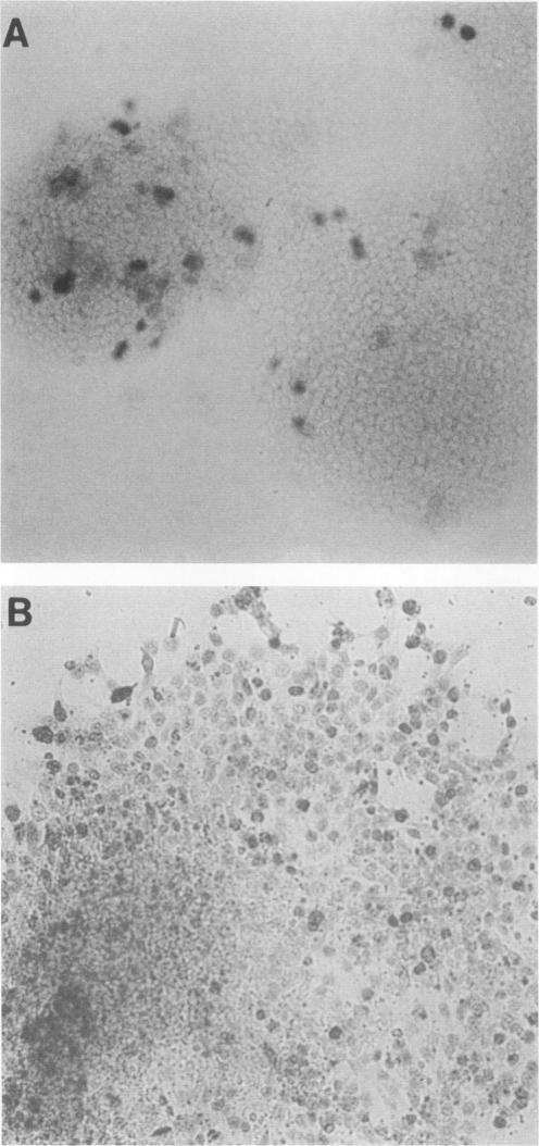
Undifferentiated F9 cells transfected with plasmids encoding adenovirus E1a gene products underwent radical morphological changes. They ceased to express the SSEA-1 stem cell marker antigen and started to express a number of the characteristics of the differentiated state that is induced in F9 cells by treatment with retinoic acid. In particular, they expressed keratin intermediate filaments and acquired the ability to synthesise simian virus 40 tumor antigens after virus infection. The transfected cells expressed the E1a proteins, and this expression was necessary to induce the phenotypic changes, since a coisogenic plasmid encoding only a truncated 70-amino-acid E1a polypeptide and the transfection procedure itself did not detectably after the morphology or marker expression of the F9 stem cells. The phenotypic change was induced by both 13S and 12S cDNA plasmids. We discuss these results in the context of known E1a functions and with reference to the other oncogenes and external factors that can cause F9 cell differentiation.
用编码腺病毒E1a基因产物的质粒转染未分化的F9细胞后,细胞发生了显著的形态变化。它们不再表达SSEA-1干细胞标记抗原,并开始表现出一些经视黄酸处理诱导F9细胞分化后所具有的分化状态特征。特别是,它们表达角蛋白中间丝,并在病毒感染后获得了合成猿猴病毒40肿瘤抗原的能力。转染细胞表达E1a蛋白,这种表达对于诱导表型变化是必需的,因为一个仅编码截短的70个氨基酸的E1a多肽的同基因质粒以及转染过程本身在F9干细胞的形态或标记表达上没有可检测到的影响。13S和12S cDNA质粒均可诱导表型变化。我们结合已知的E1a功能,并参考其他可导致F9细胞分化的癌基因和外部因素来讨论这些结果。