Jiang Hong-Ke, Wang You-Hua, Sun Lei, He Xi, Zhao Mei, Feng Zhi-Hui, Yu Xiao-Jiang, Zang Wei-Jin
Department of Pharmacology, College of Medicine, Xi'an Jiaotong University, Xi'an 710061, China.
Center for Mitochondrial Biology and Medicine, Key Laboratory of Biomedical Information Engineering of the Ministry of Education, School of Life Science and Technology, Frontier Institute of Science and Technology, Xi'an Jiaotong University, Xi'an 710049, China.
Int J Mol Sci. 2014 Mar 26;15(4):5304-22. doi: 10.3390/ijms15045304.
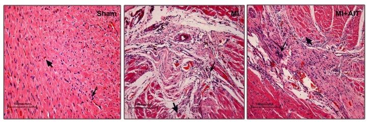
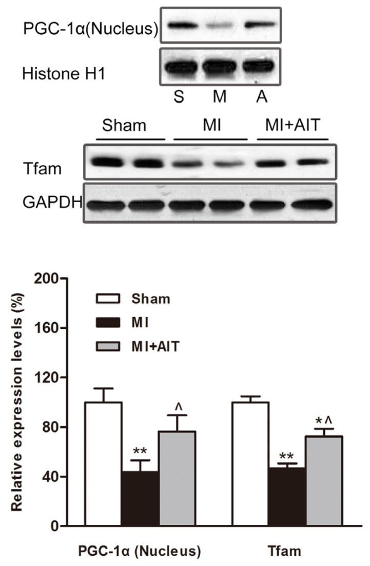
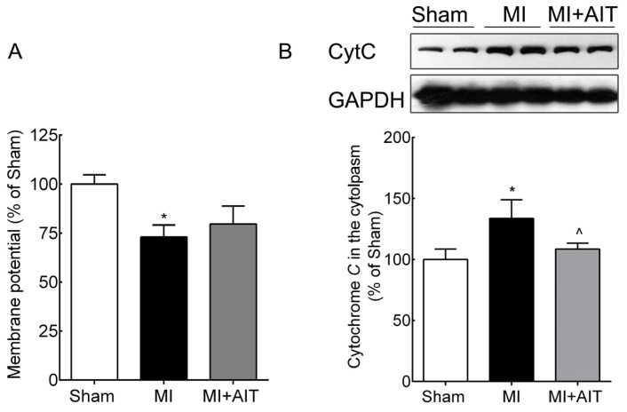
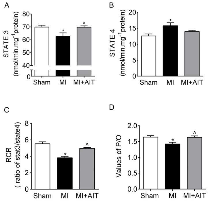
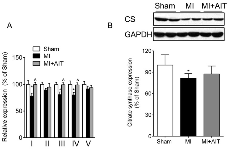
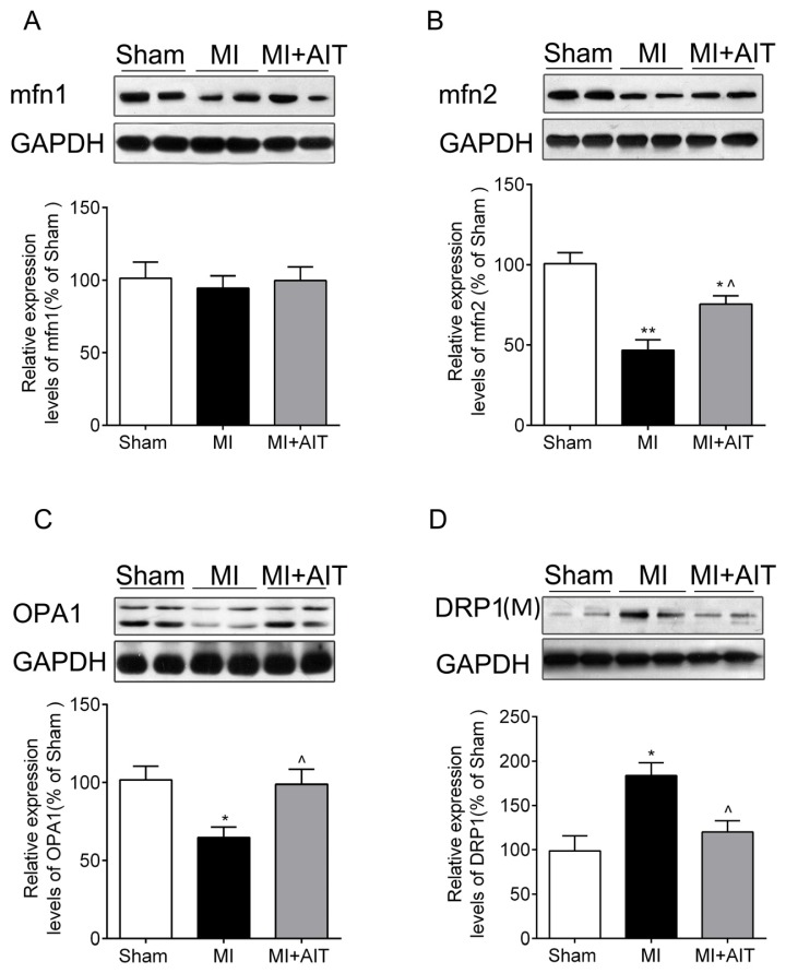
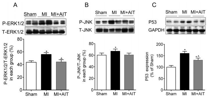
Aerobic interval training (AIT) can favorably affect cardiovascular diseases. However, the effects of AIT on post-myocardial infarction (MI)-associated mitochondrial dysfunctions remain unclear. In this study, we investigated the protective effects of AIT on myocardial mitochondria in post-MI rats by focusing on mitochondrial dynamics (fusion and fission). Mitochondrial respiratory functions (as measured by the respiratory control ratio (RCR) and the ratio of ADP to oxygen consumption (P/O)); complex activities; dynamic proteins (mitofusin (mfn) 1/2, type 1 optic atrophy (OPA1) and dynamin-related protein1 (DRP1)); nuclear peroxisome proliferator-activated receptor gamma coactivator 1-alpha (PGC-1α); and the oxidative signaling of extracellular signal-regulated kinase (ERK) 1/2, c-Jun NH2-terminal protein kinase (JNK) and P53 were observed. Post-MI rats exhibited mitochondrial dysfunction and adverse mitochondrial network dynamics (reduced fusion and increased fission), which was associated with activated ERK1/2-JNK-P53 signaling and decreased nuclear PGC-1α. After AIT, MI-associated mitochondrial dysfunction was improved (elevated RCR and P/O and enhanced complex I, III and IV activities); in addition, increased fusion (mfn2 and OPA1), decreased fission (DRP1), elevated nuclear PGC-1α and inactivation of the ERK1/2-JNK-P53 signaling were observed. These data demonstrate that AIT may restore the post-MI mitochondrial function by inhibiting dynamics pathological remodeling, which may be associated with inactivation of ERK1/2-JNK-P53 signaling and increase in nuclear PGC-1α expression.
有氧间歇训练(AIT)可对心血管疾病产生有益影响。然而,AIT对心肌梗死后(MI)相关线粒体功能障碍的影响仍不清楚。在本研究中,我们通过关注线粒体动力学(融合与裂变)来研究AIT对MI后大鼠心肌线粒体的保护作用。观察了线粒体呼吸功能(通过呼吸控制率(RCR)和ADP与氧消耗比(P/O)来衡量)、复合体活性、动态蛋白(线粒体融合蛋白(mfn)1/2、视神经萎缩蛋白1(OPA1)和动力相关蛋白1(DRP1))、核过氧化物酶体增殖物激活受体γ共激活因子1α(PGC-1α)以及细胞外信号调节激酶(ERK)1/2、c-Jun氨基末端蛋白激酶(JNK)和P53的氧化信号。MI后大鼠表现出线粒体功能障碍和不良的线粒体网络动力学(融合减少和裂变增加),这与ERK1/2-JNK-P53信号激活和核PGC-1α降低有关。AIT后,MI相关的线粒体功能障碍得到改善(RCR和P/O升高以及复合体I、III和IV活性增强);此外,观察到融合增加(mfn2和OPA1)、裂变减少(DRP1)、核PGC-1α升高以及ERK1/2-JNK-P53信号失活。这些数据表明,AIT可能通过抑制动力学病理重塑来恢复MI后的线粒体功能,这可能与ERK1/2-JNK-P53信号失活和核PGC-1α表达增加有关。