Chandra S, Ahmad T, Barth R F, Kabalka G W
Cornell SIMS Laboratory, Department of Biomedical Engineering, Cornell University, Ithaca, New York, U.S.A.
J Microsc. 2014 Jun;254(3):146-56. doi: 10.1111/jmi.12126. Epub 2014 Mar 31.
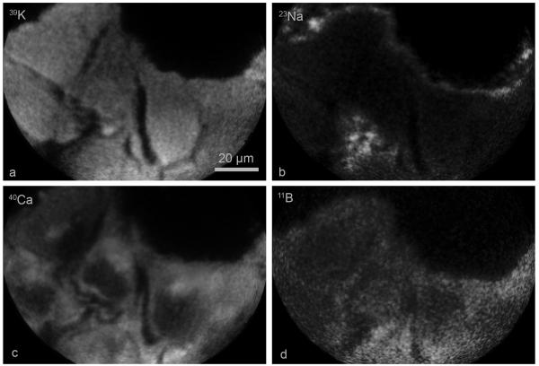
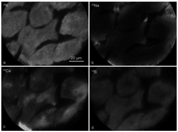
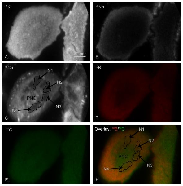
Boron neutron capture therapy (BNCT) of cancer depends on the selective delivery of a sufficient number of boron-10 ((10)B) atoms to individual tumour cells. Cell killing results from the (10)B (n, α)(7) Li neutron capture and fission reactions that occur if a sufficient number of (10)B atoms are localized in the tumour cells. Intranuclear (10)B localization enhances the efficiency of cell killing via damage to the DNA. The net cellular content of (10)B atoms reflects both bound and free pools of boron in individual tumour cells. The assessment of these pools, delivered by a boron delivery agent, currently cannot be made at subcellular-scale resolution by clinically applicable techniques such as positron emission tomography and magnetic resonance imaging. In this study, a secondary ion mass spectrometry based imaging instrument, a CAMECA IMS 3f ion microscope, capable of 500 nm spatial resolution was employed. Cryogenically prepared cultured human T98G glioblastoma cells were evaluated for boron uptake and retention of two delivery agents. The first, L-p-boronophenylalanine (BPA), has been used clinically for BNCT of high-grade gliomas, recurrent tumours of the head and neck region and melanomas. The second, a boron analogue of an unnatural amino acid, 1-amino-3-borono-cyclopentanecarboxylic acid (cis-ABCPC), has been studied in rodent glioma and melanoma models by quantification of boron in the nucleus and cytoplasm of individual tumour cells. The bound and free pools of boron were assessed by exposure of cells to boron-free nutrient medium. Both BPA and cis-ABCPC delivered almost 70% of the pool of boron in the free or loosely bound form to the nucleus and cytoplasm of human glioblastoma cells. This free pool of boron could be easily mobilized out of the cell and was in some sort of equilibrium with extracellular boron. In the case of BPA, the intracellular free pool of boron also was affected by the presence of phenylalanine in the nutrient medium. This suggests that it might be advantageous if patients were placed on a low phenylalanine diet prior to the initiation of BNCT. Since BPA currently is used clinically for BNCT, our observations may have direct relevance to future clinical studies utilizing this agent and provides support for individualized treatment planning regimens rather than the use of fixed BPA infusion protocols.
癌症的硼中子俘获疗法(BNCT)依赖于将足够数量的硼 - 10(¹⁰B)原子选择性地递送至各个肿瘤细胞。如果足够数量的¹⁰B原子定位于肿瘤细胞中,细胞杀伤则源于¹⁰B(n,α)⁷Li中子俘获和裂变反应。核内¹⁰B定位通过对DNA的损伤提高细胞杀伤效率。¹⁰B原子的净细胞含量反映了各个肿瘤细胞中硼的结合池和游离池。目前,通过正电子发射断层扫描和磁共振成像等临床适用技术,无法在亚细胞尺度分辨率下评估由硼递送剂递送的这些池。在本研究中,使用了基于二次离子质谱的成像仪器,即具有500 nm空间分辨率的CAMECA IMS 3f离子显微镜。对低温制备的培养人T98G胶质母细胞瘤细胞进行了两种递送剂的硼摄取和保留评估。第一种是L - 对硼苯丙氨酸(BPA),已在临床上用于高级别胶质瘤、头颈部复发性肿瘤和黑色素瘤的BNCT。第二种是一种非天然氨基酸的硼类似物,1 - 氨基 - 3 - 硼代环戊烷羧酸(顺式 - ABCPC),已在啮齿动物胶质瘤和黑色素瘤模型中通过对单个肿瘤细胞核和细胞质中的硼进行定量研究。通过将细胞暴露于无硼营养培养基中来评估硼的结合池和游离池。BPA和顺式 - ABCPC都将几乎70%的游离或松散结合形式的硼池递送至人胶质母细胞瘤细胞的细胞核和细胞质。这种游离硼池可以很容易地从细胞中动员出来,并与细胞外硼处于某种平衡状态。就BPA而言,营养培养基中苯丙氨酸的存在也会影响细胞内硼的游离池。这表明如果患者在BNCT开始前采用低苯丙氨酸饮食可能是有利的。由于BPA目前在临床上用于BNCT,我们的观察结果可能与未来使用该药物的临床研究直接相关,并为个体化治疗计划方案而非固定的BPA输注方案的使用提供支持。