• 文献检索
  • 文档翻译
  • 深度研究
  • 学术资讯
  • Suppr Zotero 插件Zotero 插件
  • 邀请有礼
  • 套餐&价格
  • 历史记录
应用&插件
Suppr Zotero 插件Zotero 插件浏览器插件Mac 客户端Windows 客户端微信小程序
定价
高级版会员购买积分包购买API积分包
服务
文献检索文档翻译深度研究API 文档MCP 服务
关于我们
关于 Suppr公司介绍联系我们用户协议隐私条款
关注我们

Suppr 超能文献

核心技术专利:CN118964589B侵权必究
粤ICP备2023148730 号-1Suppr @ 2026

文献检索

告别复杂PubMed语法,用中文像聊天一样搜索,搜遍4000万医学文献。AI智能推荐,让科研检索更轻松。

立即免费搜索

文件翻译

保留排版,准确专业,支持PDF/Word/PPT等文件格式,支持 12+语言互译。

免费翻译文档

深度研究

AI帮你快速写综述,25分钟生成高质量综述,智能提取关键信息,辅助科研写作。

立即免费体验

搏动性耳鸣患者的基线脑活动异常:一项静息态 fMRI 研究。

Abnormal baseline brain activity in patients with pulsatile tinnitus: a resting-state FMRI study.

机构信息

Department of Radiology Center, Beijing Tongren Hospital, Capital Medical University, Beijing 100730, China.

Department of Radiology Center, Beijing Friendship Hospital, Capital Medical University, Beijing 100050, China.

出版信息

Neural Plast. 2014;2014:549162. doi: 10.1155/2014/549162. Epub 2014 Apr 24.

DOI:10.1155/2014/549162
PMID:24872895
原文链接:https://pmc.ncbi.nlm.nih.gov/articles/PMC4020302/
Abstract

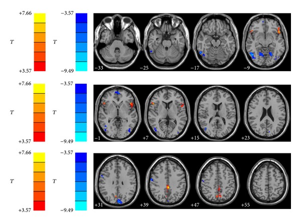
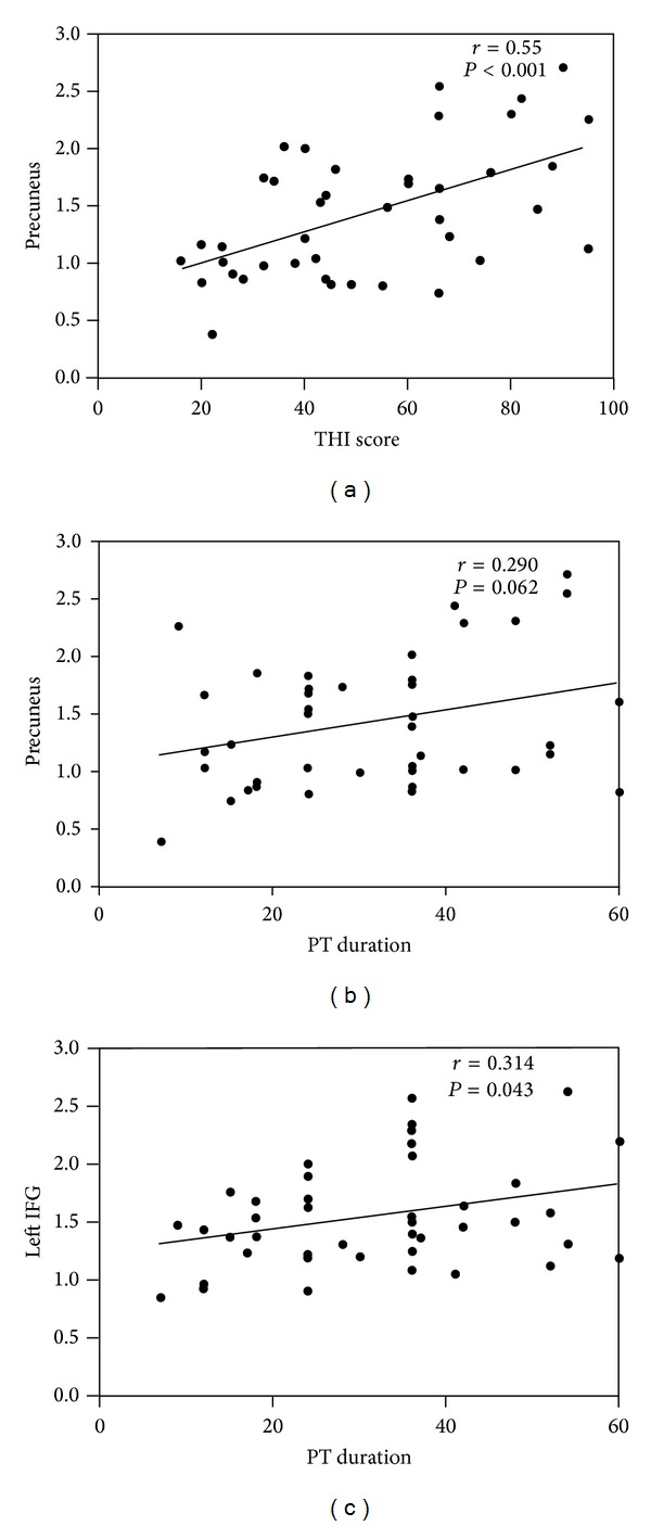
Numerous investigations studying the brain functional activity of the tinnitus patients have indicated that neurological changes are important findings of this kind of disease. However, the pulsatile tinnitus (PT) patients were excluded in previous studies because of the totally different mechanisms of the two subtype tinnitus. The aim of this study is to investigate whether altered baseline brain activity presents in patients with PT using resting-state functional magnetic resonance imaging (rs-fMRI) technique. The present study used unilateral PT patients (n = 42) and age-, sex-, and education-matched normal control subjects (n = 42) to investigate the changes in structural and amplitude of low-frequency (ALFF) of the brain. Also, we analyzed the relationships between these changes with clinical data of the PT patients. Compared with normal controls, PT patients did not show any structural changes. PT patients showed significant increased ALFF in the bilateral precuneus, and bilateral inferior frontal gyrus (IFG) and decreased ALFF in multiple occipital areas. Moreover, the increased THI score and PT duration was correlated with increased ALFF in precuneus and bilateral IFG. The abnormalities of spontaneous brain activity reflected by ALFF measurements in the absence of structural changes may provide insights into the neural reorganization in PT patients.

摘要

大量研究表明,耳鸣患者的大脑功能活动发生了改变,这是该疾病的重要发现。然而,由于两种耳鸣亚型的发病机制完全不同,以前的研究都排除了搏动性耳鸣(PT)患者。本研究旨在使用静息态功能磁共振成像(rs-fMRI)技术,探讨 PT 患者是否存在静息态脑活动改变。本研究使用单侧 PT 患者(n = 42)和年龄、性别、教育程度匹配的正常对照组(n = 42),分析脑结构和低频振幅(ALFF)的变化。此外,我们还分析了这些变化与 PT 患者临床数据之间的关系。与正常对照组相比,PT 患者的脑结构没有任何变化。PT 患者双侧楔前叶、双侧额下回(IFG)的 ALFF 显著增加,多个枕叶区域的 ALFF 减少。此外,THI 评分和 PT 持续时间的增加与楔前叶和双侧 IFG 的 ALFF 增加相关。ALFF 测量所反映的自发性脑活动异常,可能为 PT 患者的神经重组提供了新的视角。

https://cdn.ncbi.nlm.nih.gov/pmc/blobs/6a04/4020302/b215002feb0e/NP2014-549162.002.jpg
https://cdn.ncbi.nlm.nih.gov/pmc/blobs/6a04/4020302/f674b19c7cdb/NP2014-549162.001.jpg
https://cdn.ncbi.nlm.nih.gov/pmc/blobs/6a04/4020302/b215002feb0e/NP2014-549162.002.jpg
https://cdn.ncbi.nlm.nih.gov/pmc/blobs/6a04/4020302/f674b19c7cdb/NP2014-549162.001.jpg
https://cdn.ncbi.nlm.nih.gov/pmc/blobs/6a04/4020302/b215002feb0e/NP2014-549162.002.jpg

相似文献

1
Abnormal baseline brain activity in patients with pulsatile tinnitus: a resting-state FMRI study.搏动性耳鸣患者的基线脑活动异常:一项静息态 fMRI 研究。
Neural Plast. 2014;2014:549162. doi: 10.1155/2014/549162. Epub 2014 Apr 24.
2
Disrupted neural activity in unilateral vascular pulsatile tinnitus patients in the early stage of disease: evidence from resting-state fMRI.单侧血管搏动性耳鸣患者疾病早期的神经活动紊乱:静息态功能磁共振成像证据
Prog Neuropsychopharmacol Biol Psychiatry. 2015 Jun 3;59:91-99. doi: 10.1016/j.pnpbp.2015.01.013. Epub 2015 Jan 30.
3
Frequency-Dependent Neural Activity in Patients with Unilateral Vascular Pulsatile Tinnitus.单侧血管搏动性耳鸣患者的频率依赖性神经活动
Neural Plast. 2016;2016:4918186. doi: 10.1155/2016/4918186. Epub 2016 Jun 20.
4
Abnormal resting-state functional connectivity study in unilateral pulsatile tinnitus patients with single etiology: A seed-based functional connectivity study.单病因单侧搏动性耳鸣患者静息态功能连接异常研究:基于种子点的功能连接研究。
Eur J Radiol. 2016 Nov;85(11):2023-2029. doi: 10.1016/j.ejrad.2016.09.011. Epub 2016 Sep 13.
5
Resting-state functional connectivity density mapping of etiology confirmed unilateral pulsatile tinnitus patients: Altered functional hubs in the early stage of disease.病因确诊的单侧搏动性耳鸣患者静息态功能连接密度图谱:疾病早期功能枢纽的改变
Neuroscience. 2015 Dec 3;310:27-37. doi: 10.1016/j.neuroscience.2015.09.032. Epub 2015 Sep 16.
6
Aberrant spontaneous brain activity in chronic tinnitus patients revealed by resting-state functional MRI.静息态功能磁共振成像揭示慢性耳鸣患者的异常自发脑活动。
Neuroimage Clin. 2014 Sep 19;6:222-8. doi: 10.1016/j.nicl.2014.09.011. eCollection 2014.
7
Frequency-specific alternations in the amplitude of low-frequency fluctuations in chronic tinnitus.慢性耳鸣低频波动幅度的频率特异性改变
Front Neural Circuits. 2015 Oct 29;9:67. doi: 10.3389/fncir.2015.00067. eCollection 2015.
8
Abnormal regional activity and functional connectivity in resting-state brain networks associated with etiology confirmed unilateral pulsatile tinnitus in the early stage of disease.与病因相关的静息态脑网络中异常的区域活动和功能连接在疾病早期证实了单侧搏动性耳鸣。
Hear Res. 2017 Mar;346:55-61. doi: 10.1016/j.heares.2017.02.004. Epub 2017 Feb 7.
9
Surface-Based Amplitude of Low-Frequency Fluctuation Alterations in Patients With Tinnitus Before and After Sound Therapy: A Resting-State Functional Magnetic Resonance Imaging Study.耳鸣患者声音治疗前后基于表面的低频波动幅度变化:一项静息态功能磁共振成像研究
Front Neurosci. 2021 Nov 17;15:709482. doi: 10.3389/fnins.2021.709482. eCollection 2021.
10
Altered brain functional connectivity and correlation with psychological status in patients with unilateral pulsatile tinnitus.单侧搏动性耳鸣患者脑功能连接和与心理状态的相关性改变。
Neurosci Lett. 2019 Jul 13;705:235-245. doi: 10.1016/j.neulet.2019.04.046. Epub 2019 Apr 28.

引用本文的文献

1
Distinct changes in the morphology of cortical and subcortical grey matter associated with age-related hearing loss and tinnitus in the UK Biobank participants.英国生物银行参与者中,与年龄相关性听力损失和耳鸣相关的皮质及皮质下灰质形态的明显变化。
Brain Commun. 2025 May 27;7(3):fcaf203. doi: 10.1093/braincomms/fcaf203. eCollection 2025.
2
Disruptions of default mode network and precuneus connectivity associated with cognitive dysfunctions in tinnitus.耳鸣患者认知功能障碍与默认模式网络和楔前叶连接中断有关。
Sci Rep. 2023 Apr 7;13(1):5746. doi: 10.1038/s41598-023-32599-0.
3
Invariant structural and functional brain regions associated with tinnitus: A meta-analysis.

本文引用的文献

1
Epileptic discharges affect the default mode network--FMRI and intracerebral EEG evidence.癫痫放电影响默认模式网络——fMRI 和颅内 EEG 证据。
PLoS One. 2013 Jun 28;8(6):e68038. doi: 10.1371/journal.pone.0068038. Print 2013.
2
Brain regions responsible for tinnitus distress and loudness: a resting-state FMRI study.负责耳鸣困扰和响度的大脑区域:一项静息态 fMRI 研究。
PLoS One. 2013 Jun 25;8(6):e67778. doi: 10.1371/journal.pone.0067778. Print 2013.
3
The Quest for EEG Power Band Correlation with ICA Derived fMRI Resting State Networks.
与耳鸣相关的不变结构和功能脑区:一项荟萃分析。
PLoS One. 2022 Oct 18;17(10):e0276140. doi: 10.1371/journal.pone.0276140. eCollection 2022.
4
Altered cerebral blood flow in patients with unilateral venous pulsatile tinnitus: an arterial spin labeling study.单侧静脉性搏动性耳鸣患者脑血流改变:一项动脉自旋标记研究。
Br J Radiol. 2021 Apr 1;94(1120):20200990. doi: 10.1259/bjr.20200990. Epub 2021 Mar 18.
5
Lateralization Effects on Cerebral Blood Flow in Patients With Unilateral Pulsatile Tinnitus Measured With Arterial Spin Labeling.动脉自旋标记法测量单侧搏动性耳鸣患者脑血流的偏侧化效应
Front Hum Neurosci. 2020 Nov 17;14:591260. doi: 10.3389/fnhum.2020.591260. eCollection 2020.
6
Disrupted Intraregional Brain Activity and Functional Connectivity in Unilateral Acute Tinnitus Patients With Hearing Loss.单侧急性耳鸣伴听力损失患者的脑区内部活动和功能连接中断
Front Neurosci. 2019 Sep 19;13:1010. doi: 10.3389/fnins.2019.01010. eCollection 2019.
7
Tinnitus distress is associated with enhanced resting-state functional connectivity within the default mode network.耳鸣困扰与默认模式网络内静息态功能连接增强有关。
Neuropsychiatr Dis Treat. 2018 Aug 1;14:1919-1927. doi: 10.2147/NDT.S164619. eCollection 2018.
8
Neuroanatomical Alterations in Patients with Early Stage of Unilateral Pulsatile Tinnitus: A Voxel-Based Morphometry Study.单侧搏动性耳鸣早期患者的神经解剖结构改变:基于体素的形态测量学研究。
Neural Plast. 2018 Feb 28;2018:4756471. doi: 10.1155/2018/4756471. eCollection 2018.
9
Connectivity of precuneus to the default mode and dorsal attention networks: A possible invariant marker of long-term tinnitus.楔前叶与默认模式网络和背侧注意网络的连接性:长期耳鸣的一种可能的不变标志物。
Neuroimage Clin. 2017 Jul 22;16:196-204. doi: 10.1016/j.nicl.2017.07.015. eCollection 2017.
10
Frequency-Dependent Neural Activity in Patients with Unilateral Vascular Pulsatile Tinnitus.单侧血管搏动性耳鸣患者的频率依赖性神经活动
Neural Plast. 2016;2016:4918186. doi: 10.1155/2016/4918186. Epub 2016 Jun 20.
探索 EEG 功率带与基于 ICA 的 fMRI 静息态网络的相关性。
Front Hum Neurosci. 2013 Jun 25;7:315. doi: 10.3389/fnhum.2013.00315. eCollection 2013.
4
Brain areas controlling heart rate variability in tinnitus and tinnitus-related distress.控制耳鸣和耳鸣相关困扰的心率变异性的大脑区域。
PLoS One. 2013;8(3):e59728. doi: 10.1371/journal.pone.0059728. Epub 2013 Mar 22.
5
Auditory cortex is implicated in tinnitus distress: a voxel-based morphometry study.听觉皮层与耳鸣困扰有关:基于体素的形态测量学研究。
Brain Struct Funct. 2013 Jul;218(4):1061-70. doi: 10.1007/s00429-013-0520-z. Epub 2013 Feb 24.
6
Speech-induced striatal dopamine release is left lateralized and coupled to functional striatal circuits in healthy humans: a combined PET, fMRI and DTI study.健康人类中,语言诱导纹状体多巴胺释放具有左侧偏侧化特征,并与功能性纹状体回路相关:一项结合 PET、fMRI 和 DTI 的研究。
Neuroimage. 2013 Apr 15;70:21-32. doi: 10.1016/j.neuroimage.2012.12.042. Epub 2012 Dec 28.
7
A default mode of brain function in altered states of consciousness.意识改变状态下大脑功能的一种默认模式。
Arch Ital Biol. 2012 Jun-Sep;150(2-3):107-21. doi: 10.4449/aib.v150i2.1373.
8
Functional MRI evidence for a role of ventral prefrontal cortex in tinnitus.功能性磁共振成像证据表明,腹侧前额叶皮层在耳鸣中起作用。
Brain Res. 2012 Nov 16;1485:22-39. doi: 10.1016/j.brainres.2012.08.052. Epub 2012 Sep 6.
9
The auditory and non-auditory brain areas involved in tinnitus. An emergent property of multiple parallel overlapping subnetworks.涉及耳鸣的听觉和非听觉脑区。多个并行重叠子网的新兴特性。
Front Syst Neurosci. 2012 May 8;6:31. doi: 10.3389/fnsys.2012.00031. eCollection 2012.
10
Connectivity graph analysis of the auditory resting state network in tinnitus.耳鸣静息态网络的连通性图谱分析。
Brain Res. 2012 Nov 16;1485:10-21. doi: 10.1016/j.brainres.2012.05.006. Epub 2012 May 10.