• 文献检索
  • 文档翻译
  • 深度研究
  • 学术资讯
  • Suppr Zotero 插件Zotero 插件
  • 邀请有礼
  • 套餐&价格
  • 历史记录
应用&插件
Suppr Zotero 插件Zotero 插件浏览器插件Mac 客户端Windows 客户端微信小程序
定价
高级版会员购买积分包购买API积分包
服务
文献检索文档翻译深度研究API 文档MCP 服务
关于我们
关于 Suppr公司介绍联系我们用户协议隐私条款
关注我们

Suppr 超能文献

核心技术专利:CN118964589B侵权必究
粤ICP备2023148730 号-1Suppr @ 2026

文献检索

告别复杂PubMed语法,用中文像聊天一样搜索,搜遍4000万医学文献。AI智能推荐,让科研检索更轻松。

立即免费搜索

文件翻译

保留排版,准确专业,支持PDF/Word/PPT等文件格式,支持 12+语言互译。

免费翻译文档

深度研究

AI帮你快速写综述,25分钟生成高质量综述,智能提取关键信息,辅助科研写作。

立即免费体验

贝塞尔光束在活样本中的扩展双光子显微镜:更稳定的焦点,更快的体积扫描,以及更简单的立体成像。

Extended two-photon microscopy in live samples with Bessel beams: steadier focus, faster volume scans, and simpler stereoscopic imaging.

机构信息

Département de Physique, de Génie Physique et d'Optique, Centre d'Optique, Photonique et Laser, Université Laval Québec, QC, Canada ; Centre de Recherche de l'Institut Universitaire en Santé Mentale de Québec Québec, QC, Canada.

Centre de Recherche de l'Institut Universitaire en Santé Mentale de Québec Québec, QC, Canada.

出版信息

Front Cell Neurosci. 2014 May 20;8:139. doi: 10.3389/fncel.2014.00139. eCollection 2014.

DOI:10.3389/fncel.2014.00139
PMID:24904284
原文链接:https://pmc.ncbi.nlm.nih.gov/articles/PMC4032997/
Abstract

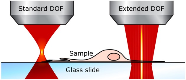
Two-photon microscopy has revolutionized functional cellular imaging in tissue, but although the highly confined depth of field (DOF) of standard set-ups yields great optical sectioning, it also limits imaging speed in volume samples and ease of use. For this reason, we recently presented a simple and retrofittable modification to the two-photon laser-scanning microscope which extends the DOF through the use of an axicon (conical lens). Here we demonstrate three significant benefits of this technique using biological samples commonly employed in the field of neuroscience. First, we use a sample of neurons grown in culture and move it along the z-axis, showing that a more stable focus is achieved without compromise on transverse resolution. Second, we monitor 3D population dynamics in an acute slice of live mouse cortex, demonstrating that faster volumetric scans can be conducted. Third, we acquire a stereoscopic image of neurons and their dendrites in a fixed sample of mouse cortex, using only two scans instead of the complete stack and calculations required by standard systems. Taken together, these advantages, combined with the ease of integration into pre-existing systems, make the extended depth-of-field imaging based on Bessel beams a strong asset for the field of microscopy and life sciences in general.

摘要

双光子显微镜技术已经彻底改变了组织中的功能细胞成像,但尽管标准设置具有高度受限的景深(DOF),能够实现出色的光学切片,但它也限制了体积样本的成像速度和易用性。出于这个原因,我们最近对双光子激光扫描显微镜进行了简单的改装和改造,通过使用轴棱锥(锥形透镜)来扩展景深。在这里,我们使用神经科学领域常用的生物样本展示了这项技术的三个重要优势。首先,我们使用培养的神经元样本在 z 轴上移动,结果表明,在不影响横向分辨率的情况下,可以实现更稳定的焦点。其次,我们在活体小鼠皮层的急性切片中监测 3D 群体动力学,证明可以进行更快的体积扫描。第三,我们使用仅两次扫描而不是标准系统所需的完整堆栈和计算,从固定的小鼠皮层样本中获取神经元及其树突的立体图像。总之,这些优势,加上易于集成到现有系统中,使得基于贝塞尔光束的扩展景深成像成为显微镜和生命科学领域的有力工具。

https://cdn.ncbi.nlm.nih.gov/pmc/blobs/9d7f/4032997/de2601d39494/fncel-08-00139-g0009.jpg
https://cdn.ncbi.nlm.nih.gov/pmc/blobs/9d7f/4032997/1c9ba666d375/fncel-08-00139-g0001.jpg
https://cdn.ncbi.nlm.nih.gov/pmc/blobs/9d7f/4032997/6537edd11bf9/fncel-08-00139-g0002.jpg
https://cdn.ncbi.nlm.nih.gov/pmc/blobs/9d7f/4032997/41b51a3f3bba/fncel-08-00139-g0003.jpg
https://cdn.ncbi.nlm.nih.gov/pmc/blobs/9d7f/4032997/81e1584fcc6b/fncel-08-00139-g0004.jpg
https://cdn.ncbi.nlm.nih.gov/pmc/blobs/9d7f/4032997/118c6b19a828/fncel-08-00139-g0005.jpg
https://cdn.ncbi.nlm.nih.gov/pmc/blobs/9d7f/4032997/ba54442b588b/fncel-08-00139-g0006.jpg
https://cdn.ncbi.nlm.nih.gov/pmc/blobs/9d7f/4032997/a2f683d4f48f/fncel-08-00139-g0007.jpg
https://cdn.ncbi.nlm.nih.gov/pmc/blobs/9d7f/4032997/fab2e3758470/fncel-08-00139-g0008.jpg
https://cdn.ncbi.nlm.nih.gov/pmc/blobs/9d7f/4032997/de2601d39494/fncel-08-00139-g0009.jpg

相似文献

1
Extended two-photon microscopy in live samples with Bessel beams: steadier focus, faster volume scans, and simpler stereoscopic imaging.贝塞尔光束在活样本中的扩展双光子显微镜:更稳定的焦点,更快的体积扫描,以及更简单的立体成像。
Front Cell Neurosci. 2014 May 20;8:139. doi: 10.3389/fncel.2014.00139. eCollection 2014.
2
Rapid, high-contrast, and steady volumetric imaging with Bessel-beam-based two-photon fluorescence microscopy.基于贝塞尔光束的双光子荧光显微镜的快速、高对比度、稳定的体积成像。
J Biomed Opt. 2024 Jan;29(1):016501. doi: 10.1117/1.JBO.29.1.016501. Epub 2024 Jan 24.
3
Rapid volumetric imaging with Bessel-Beam three-photon microscopy.利用贝塞尔光束三光子显微镜进行快速容积成像。
Biomed Opt Express. 2018 Mar 29;9(4):1992-2000. doi: 10.1364/BOE.9.001992. eCollection 2018 Apr 1.
4
Two-photon scanned light sheet fluorescence microscopy with axicon imaging for fast volumetric imaging.双光子扫描光片荧光显微镜结合轴棱锥成像用于快速体积成像。
J Biomed Opt. 2021 Nov;26(11). doi: 10.1117/1.JBO.26.11.116503.
5
Resolution enhancement in an extended depth of field for volumetric two-photon microscopy.体积双光子显微镜扩展景深中的分辨率增强。
Opt Lett. 2020 Jun 1;45(11):3054-3057. doi: 10.1364/OL.394282.
6
Wide field light-sheet microscopy with lens-axicon controlled two-photon Bessel beam illumination.广角光片显微镜与透镜-轴棱锥控制双光子贝塞尔光束照明。
Nat Commun. 2021 May 20;12(1):2979. doi: 10.1038/s41467-021-23249-y.
7
Image scanning microscopy with multiphoton excitation or Bessel beam illumination.采用多光子激发或贝塞尔光束照明的图像扫描显微镜。
J Opt Soc Am A Opt Image Sci Vis. 2020 Oct 1;37(10):1639-1649. doi: 10.1364/JOSAA.402048.
8
Depth random-access two-photon Bessel light-sheet imaging in brain tissue.脑组织中深度随机访问双光子贝塞尔光片成像。
Opt Express. 2022 Jul 18;30(15):26396-26406. doi: 10.1364/OE.456871.
9
Bessel beam spectral-domain high-resolution optical coherence tomography with micro-optic axicon providing extended focusing range.采用微光学轴棱锥实现扩展聚焦范围的贝塞尔光束光谱域高分辨率光学相干断层扫描技术。
Opt Lett. 2008 Aug 1;33(15):1696-8. doi: 10.1364/ol.33.001696.
10
Millimetric scale two-photon Bessel-Gauss beam light sheet microscopy with three-axis isotropic resolution using an axicon lens.使用轴棱锥透镜的具有三轴各向同性分辨率的毫米级尺度双光子贝塞尔-高斯光束光片显微镜
Neurophotonics. 2023 Jul;10(3):035002. doi: 10.1117/1.NPh.10.3.035002. Epub 2023 Jun 23.

引用本文的文献

1
Modal focal adaptive optics for Bessel-focus two-photon fluorescence microscopy.用于贝塞尔聚焦双光子荧光显微镜的模态聚焦自适应光学
Opt Express. 2025 Jan 13;33(1):680-693. doi: 10.1364/OE.541033.
2
Extended-depth of field random illumination microscopy, EDF-RIM, provides super-resolved projective imaging.扩展景深随机照明显微镜(EDF-RIM)提供超分辨投影成像。
Light Sci Appl. 2024 Oct 10;13(1):285. doi: 10.1038/s41377-024-01612-0.
3
High speed two-photon laser scanning stereomicroscopy for three-dimension tracking multiple particles simultaneously in three-dimension.

本文引用的文献

1
Extended depth of field microscopy for rapid volumetric two-photon imaging.用于快速体积双光子成像的扩展景深显微镜术
Opt Express. 2013 Apr 22;21(8):10095-104. doi: 10.1364/OE.21.010095.
2
Needles of longitudinally polarized light: guidelines for minimum spot size and tunable axial extent.纵向偏振光针:最小光斑尺寸和可调轴向范围指南。
Opt Express. 2012 Jul 2;20(14):14891-905. doi: 10.1364/OE.20.014891.
3
Aberration-free three-dimensional multiphoton imaging of neuronal activity at kHz rates.无像差三维多光子成像技术以 kHz 速率对神经元活动进行成像。
用于在三维空间中同时三维跟踪多个粒子的高速双光子激光扫描立体显微镜。
Front Photon. 2022;3. doi: 10.3389/fphot.2022.985474. Epub 2022 Sep 6.
4
Rapid, high-contrast, and steady volumetric imaging with Bessel-beam-based two-photon fluorescence microscopy.基于贝塞尔光束的双光子荧光显微镜的快速、高对比度、稳定的体积成像。
J Biomed Opt. 2024 Jan;29(1):016501. doi: 10.1117/1.JBO.29.1.016501. Epub 2024 Jan 24.
5
Optical-resolution photoacoustic microscopy with a needle-shaped beam.具有针状光束的光学分辨率光声显微镜。
Nat Photonics. 2023 Jan;17(1):89-95. doi: 10.1038/s41566-022-01112-w. Epub 2022 Dec 1.
6
Two-photon calcium imaging of neuronal activity.神经元活动的双光子钙成像
Nat Rev Methods Primers. 2022;2(1). doi: 10.1038/s43586-022-00147-1. Epub 2022 Sep 1.
7
Dynamic Volumetric Imaging of Mouse Cerebral Blood Vessels In Vivo with an Ultralong Anti-Diffracting Beam.利用超长消衍射光束对活体小鼠脑血管进行动态容积成像
Molecules. 2023 Jun 23;28(13):4936. doi: 10.3390/molecules28134936.
8
Three-photon excited fluorescence imaging in neuroscience: From principles to applications.神经科学中的三光子激发荧光成像:从原理到应用
Front Neurosci. 2023 Feb 20;17:1085682. doi: 10.3389/fnins.2023.1085682. eCollection 2023.
9
Review of data processing of functional optical microscopy for neuroscience.神经科学中功能性光学显微镜的数据处理综述。
Neurophotonics. 2022 Oct;9(4):041402. doi: 10.1117/1.NPh.9.4.041402. Epub 2022 Aug 4.
10
Advantages, Pitfalls, and Developments of All Optical Interrogation Strategies of Microcircuits .微电路全光询问策略的优势、陷阱与发展
Front Neurosci. 2022 Jun 28;16:859803. doi: 10.3389/fnins.2022.859803. eCollection 2022.
Proc Natl Acad Sci U S A. 2012 Feb 21;109(8):2919-24. doi: 10.1073/pnas.1111662109. Epub 2012 Feb 6.
4
Fast two-photon in vivo imaging with three-dimensional random-access scanning in large tissue volumes.快速双光子在大组织体积中的三维随机存取扫描的体内成像。
Nat Methods. 2012 Jan 8;9(2):201-8. doi: 10.1038/nmeth.1851.
5
Efficacy of synaptic inhibition depends on multiple, dynamically interacting mechanisms implicated in chloride homeostasis.突触抑制的功效取决于多种动态相互作用的机制,这些机制与氯离子动态平衡有关。
PLoS Comput Biol. 2011 Sep;7(9):e1002149. doi: 10.1371/journal.pcbi.1002149. Epub 2011 Sep 8.
6
Deep and fast live imaging with two-photon scanned light-sheet microscopy.双光子激发扫描光片显微镜的深层快速活体成像。
Nat Methods. 2011 Jul 17;8(9):757-60. doi: 10.1038/nmeth.1652.
7
Adaptive movement compensation for in vivo imaging of fast cellular dynamics within a moving tissue.在移动组织中对快速细胞动力学进行活体成像的自适应运动补偿。
PLoS One. 2011;6(5):e19928. doi: 10.1371/journal.pone.0019928. Epub 2011 May 24.
8
Simultaneous two-photon calcium imaging at different depths with spatiotemporal multiplexing.利用时空复用技术在不同深度进行双光子钙成像。
Nat Methods. 2011 Feb;8(2):139-42. doi: 10.1038/nmeth.1552. Epub 2011 Jan 9.
9
High-speed in vivo calcium imaging reveals neuronal network activity with near-millisecond precision.高速在体钙成像以近毫秒的精度揭示神经元网络活动。
Nat Methods. 2010 May;7(5):399-405. doi: 10.1038/nmeth.1453. Epub 2010 Apr 18.
10
SLM Microscopy: Scanless Two-Photon Imaging and Photostimulation with Spatial Light Modulators.SLM 显微镜:基于空间光调制器的无扫描双光子成像和光刺激。
Front Neural Circuits. 2008 Dec 19;2:5. doi: 10.3389/neuro.04.005.2008. eCollection 2008.