Choi Keun Hee, Chung Seung Joon, Kang Min Jae, Yoon Ju Young, Lee Ji Eun, Lee Young Ah, Shin Choong Ho, Yang Sei Won
Department of Pediatrics, Seoul National University College of Medicine, Seoul, Korea.
Department of Pediatrics, Seoul National University Bundang Hospital, Seongnam, Korea.
Ann Pediatr Endocrinol Metab. 2013 Dec;18(4):183-90. doi: 10.6065/apem.2013.18.4.183. Epub 2013 Dec 31.
Brain magnetic resonance imaging (MRI) findings and factors predictive of pathological brain lesions in boys with precocious puberty (PP) or early puberty (EP) were investigated.
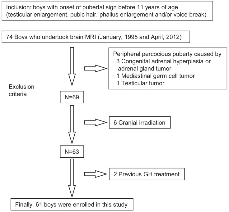
Sixty-one boys with PP or EP who had brain MRI performed were included. PP was classified into the central or peripheral type. Brain MRI findings were categorized into group I (pathological brain lesion known to cause puberty; newly diagnosed [group Ia] or previously diagnosed [group Ib]); group II (brain lesion possibly related to puberty); and group III (incidental or normal findings). Medical history, height, weight, hormone test results, and bone age were reviewed.
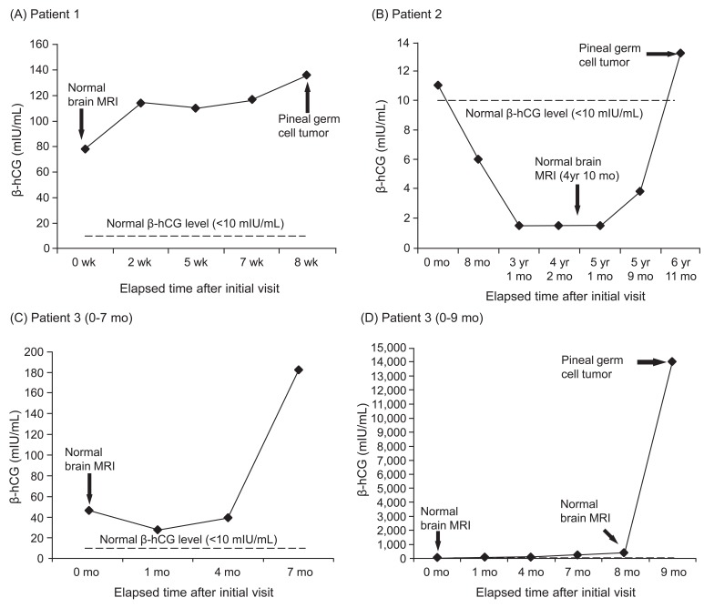
Brain lesions in groups I and II were detected in 17 of 23 boys (74%) with central PP, 9 of 30 boys (30%) with EP, and 7 of 8 boys (88%) with peripheral PP. All brain lesions in boys with peripheral PP were germ cell tumors (GCT), and 3 lesions developed later during follow-up. Group I showed earlier pubertal onset (P<0.01) and greater bone age advancement (P<0.05) than group III. Group III had lower birth weight and fewer neurological symptoms than "Ia and II" (all P<0.05).
Earlier onset of puberty, greater bone age advancement, and/or neurological symptoms suggested a greater chance of pathological brain lesions in boys with central PP or EP. All boys with peripheral PP, even those with normal initial MRI findings, should be evaluated for the emergence of GCT during follow-up.
研究性早熟(PP)或青春期提前(EP)男孩的脑磁共振成像(MRI)结果及预测病理性脑病变的因素。
纳入61例进行了脑MRI检查的PP或EP男孩。PP分为中枢性或外周性。脑MRI结果分为I组(已知可导致青春期的病理性脑病变;新诊断[Ia组]或先前已诊断[Ib组]);II组(可能与青春期相关的脑病变);III组(偶然发现或正常结果)。回顾病史、身高、体重、激素检测结果和骨龄。
23例中枢性PP男孩中有17例(74%)、30例EP男孩中有9例(30%)、8例外周性PP男孩中有7例(88%)检测到I组和II组的脑病变。外周性PP男孩的所有脑病变均为生殖细胞瘤(GCT),3例病变在随访后期出现。I组比III组青春期开始更早(P<0.01),骨龄进展更大(P<0.05)。III组出生体重较低,神经症状较“Ia组和II组”少(均P<0.05)。
青春期开始较早、骨龄进展较大和/或出现神经症状提示中枢性PP或EP男孩发生病理性脑病变的可能性更大。所有外周性PP男孩,即使初始MRI结果正常,在随访期间也应评估是否出现GCT。