Center for Neuroscience and Regenerative Medicine, Uniformed Services University of the Health Sciences , Bethesda, MD , USA ; Department of Pharmacology, Uniformed Services University of the Health Sciences , Bethesda, MD , USA.
Center for Neuroscience and Regenerative Medicine, Uniformed Services University of the Health Sciences , Bethesda, MD , USA ; Department of Anatomy, Physiology and Genetics, Uniformed Services University of the Health Sciences , Bethesda, MD , USA.
Front Neurol. 2014 Jun 4;5:82. doi: 10.3389/fneur.2014.00082. eCollection 2014.
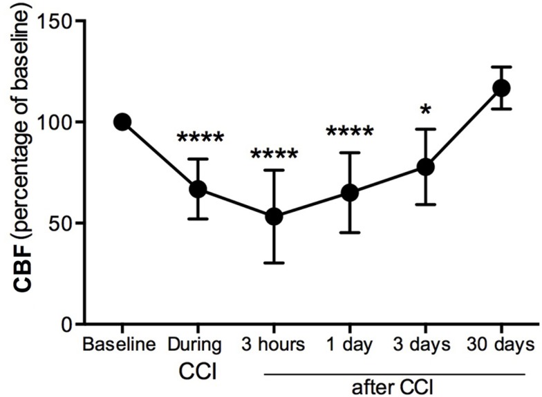
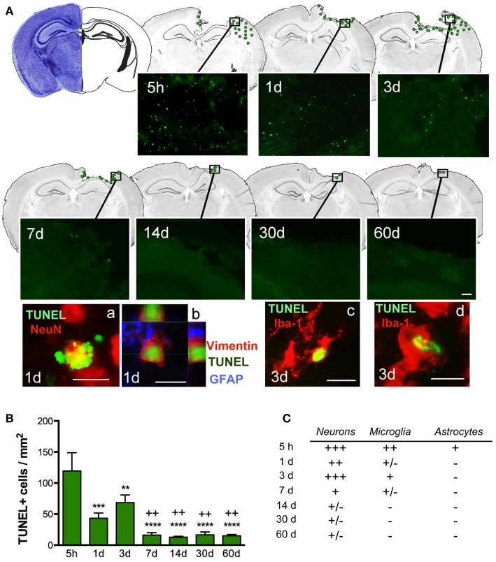
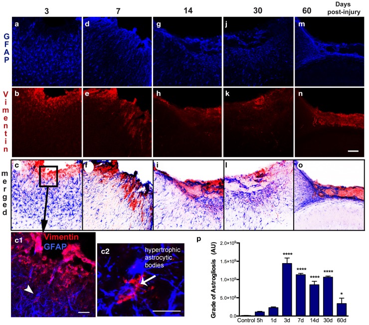
Traumatic brain injury (TBI) results in a loss of brain tissue at the moment of impact in the cerebral cortex. Subsequent secondary injury involves the release of molecular signals with dramatic consequences for the integrity of damaged tissue, leading to the evolution of a pericontusional-damaged area minutes to days after in the initial injury. The mechanisms behind the progression of tissue loss remain under investigation. In this study, we analyzed the spatial-temporal profile of blood flow, apoptotic, and astrocytic-vascular events in the cortical regions around the impact site at time points ranging from 5 h to 2 months after TBI. We performed a mild-moderate controlled cortical impact injury in young adult mice and analyzed the glial and vascular response to injury. We observed a dramatic decrease in perilesional cerebral blood flow (CBF) immediately following the cortical impact that lasted until days later. CBF finally returned to baseline levels by 30 days post-injury (dpi). The initial impact also resulted in an immediate loss of tissue and cavity formation that gradually increased in size until 3 dpi. An increase in dying cells localized in the pericontusional region and a robust astrogliosis were also observed at 3 dpi. A strong vasculature interaction with astrocytes was established at 7 dpi. Glial scar formation began at 7 dpi and seemed to be compact by 60 dpi. Altogether, these results suggest that TBI results in a progression from acute neurodegeneration that precedes astrocytic activation, reformation of the neurovascular unit to glial scar formation. Understanding the multiple processes occurring after TBI is critical to the ability to develop neuroprotective therapeutics to ameliorate the short and long-term consequences of brain injury.
创伤性脑损伤 (TBI) 导致大脑皮质在撞击瞬间失去脑组织。随后的继发性损伤涉及分子信号的释放,对受损组织的完整性产生巨大影响,导致初始损伤后几分钟到几天内伤周损伤区域的演变。组织损失进展背后的机制仍在研究中。在这项研究中,我们分析了 TBI 后 5 小时至 2 个月时间点撞击部位周围皮质区域血流、凋亡和星形胶质细胞-血管事件的时空分布。我们对年轻成年小鼠进行了轻度至中度的皮质控制冲击损伤,并分析了对损伤的神经胶质和血管反应。我们观察到皮质冲击后立即出现的伤周脑血流 (CBF) 急剧下降,这种情况持续到几天后。CBF 最终在损伤后 30 天(dpi)恢复到基线水平。最初的冲击还导致组织立即丢失和腔形成,直到 3 dpi 逐渐增大。还观察到在伤周区域定位的死亡细胞增加和强烈的星形胶质细胞增生。在 7 dpi 还观察到强烈的血管与星形胶质细胞相互作用。神经胶质瘢痕形成始于 7 dpi,到 60 dpi 时似乎已经致密。总之,这些结果表明,TBI 导致急性神经退行性变,随后是星形胶质细胞激活、神经血管单元重构到神经胶质瘢痕形成的进展。了解 TBI 后发生的多种过程对于开发神经保护治疗以改善脑损伤的短期和长期后果至关重要。