Ponnaiyan Deepa
Department of Periodontics, S.R.M Dental College and Hospital, Ramapuram, Chennai, Tamil Nadu, India.
Dent Res J (Isfahan). 2014 Mar;11(2):163-72.
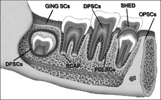
Dental tissues provide an alternate source of stem cells compared with bone marrow and have a similar potency as that of bone marrow derived mesenchymal stem cells. It has been established there are six types of dental stem cells: Dental pulp stem cells, stem cells from human exfoliated deciduous teeth, stem cells from apical papilla, periodontal ligament stem cells, dental follicle progenitor cells, oral periosteum stem cells and recently gingival connective tissue stem cells. Most of the dental tissues have a common developmental pathway; thus, it is relevant to understand whether stem cells derived from these closely related tissues are programmed differently. The present review analyzes whether stem cells form dental tissues depict distinct characteristics by gaining insight into differences in their immunophenotype. In addition, to explore the possibility of establishing a unique phenotypic fingerprint of these stem cells by identifying the unique markers that can be used to isolate these stem cells. This, in future will help in developing better techniques and markers for identification and utilization of these stem cells for regenerative therapy.
与骨髓相比,牙组织提供了另一种干细胞来源,并且具有与骨髓来源的间充质干细胞相似的潜能。现已确定有六种类型的牙干细胞:牙髓干细胞、人脱落乳牙干细胞、根尖乳头干细胞、牙周膜干细胞、牙囊祖细胞、口腔骨膜干细胞以及最近发现的牙龈结缔组织干细胞。大多数牙组织具有共同的发育途径;因此,了解源自这些密切相关组织的干细胞是否具有不同的编程方式具有重要意义。本综述通过深入研究它们免疫表型的差异,分析源自牙组织的干细胞是否具有独特特征。此外,通过鉴定可用于分离这些干细胞的独特标志物,探索建立这些干细胞独特表型指纹的可能性。这在未来将有助于开发更好的技术和标志物,用于识别和利用这些干细胞进行再生治疗。